


")
A new Tel Aviv University study combines Hasmonean history with evidence from the Dead Sea Scrolls to solve a decades-old mystery.


Research
A new Tel Aviv University study combines Hasmonean history with evidence from the Dead Sea Scrolls to solve a decades-old mystery.
A new study from Tel Aviv University proposes a solution to a historical mystery that has puzzled researchers for decades: Was the unique calendar of the Qumran sect, based on a 364-day year, ever used in practice, or was it merely a theoretical model?
The study hypothesizes that the calendar was indeed used by the sect in its early years, and even stood at the crux of the controversy that drove the sect to isolation in the desert. However, the calendar was later abandoned due to an inherent problem that made it impracticable over time, as well as warming relations with Hasmonean leadership under Alexander Jannaeus.
The study was conducted by Prof. Eshbal Ratzon of the Department of Jewish Philosophy and the Cohn Institute for the History and Philosophy of Science and Ideas at the Entin Faculty of Humanities. The paper was published in the Tarbiz Quarterly for Jewish Studies.
The Dead Sea Scrolls are a collection of ancient religious writings and fragments discovered in caves near Qumran and at other sites in the Judean Desert. Dating mainly from the Second Temple period, they include copies of biblical books as well as prayers, legal writings, commentaries, apocalyptic works, and texts describing the beliefs and practices of specific Jewish communities. The Qumran scrolls are especially important because they offer a rare window into Jewish religious life, debate, and diversity more than 2,000 years ago.

the Dead Sea Scrolls
The Qumran calendar differed from the lunisolar calendar that served as a basis for Jewish life during the Second Temple period. It consisted of exactly 364 days - a number that is perfectly divisible by seven, and thus every year included 52 full weeks, and every holiday always fell on the same day of the week. For the Qumran sect, this reflected a perfect divine order. In the political sense, the calendar represented a rebellion against the political and religious leadership at the Temple in Jerusalem, which determined all significant dates. The sect believed that these dates had already been set by God during the Creation of the world, and humans must not interfere.
Yet the Qumran calendar's mathematical perfection created a serious difficulty: it diverged by one day and a quarter from the 365-day astronomical year. This difference may appear negligible, but it accumulates rapidly. For instance, if the Qumran calendar was used for twenty years, the festivals would shift by almost four weeks relative to the seasons. After several decades, a spring festival would end up being celebrated in winter, or even in the fall. For a community that regarded festivals as agricultural events connected to the harvest, first fruits, and seasons of the year, this posed a fundamental problem.
To understand the significance of the gap, one can compare the calendar to a clock that deviates by one minute every day. At first, no one notices the problem, but after months and years, such a clock no longer represents reality. The study explains that this is exactly what happened to the Qumran calendar: while ideal from a conceptual and mathematical perspective, over time it drifted further and further away from the natural cycles it sought to govern.
Over the past decades, researchers have proposed various solutions to this enigma. Some maintained that the Qumran sect periodically added days or weeks to its calendar, while others claimed that the calendar had never actually been used in the real world, serving only as a theoretical framework. Prof. Ratzon argues that neither option is supported by the Dead Sea Scrolls. According to her study, the evidence indicates that the calendar was regarded by the sect as a key component of their religious identity and a major point of contention with the Jerusalem establishment.
The study notes that almost twenty of the scrolls found in Qumran deal with calendars and astronomy - an exceptional number that attests to the topic's immense importance for the community. The Book of Jubilees, a central work in the Qumran library, fiercely attacks the prevailing lunar calendar, presenting the 364-day calendar as the original calendar received by Moses on Mount Sinai.

Qumran caves
Based on this body of evidence, Prof. Ratzon proposes a new historical reconstruction: she contends that the Qumran calendar was actively used during the sect's formative days in the second century BCE, exacerbating its conflict with the religious leadership in Jerusalem. However, as the years went by, the calendar's accumulating digression from the seasons could no longer be ignored. In addition, the sect's relations with the ruling Hasmonean dynasty warmed during the reign of Alexander Jannaeus, who supported a halacha similar to their own and opposed the Pharisaic leadership. This enabled the Qumran sect to relinquish their previously adamant position and adopt the more practical calendar used at the Temple. They retained their own calendar as a theoretical concept that had been valid at the time of Creation and might be used again in the End of Days.
Beyond resolving a long-standing question about the Dead Sea Scrolls, the study offers insight into how religious communities respond when deeply held ideals encounter practical limitations and changing political realities. It suggests that a system can cease to function in everyday life while continuing to shape identity, belief, and collective memory.
Prof. Ratzon concludes:
"The Qumran calendar has long been regarded as one of the Qumran sect's defining features, but also as one of the most baffling mysteries of the Dead Sea Scrolls. This study proposes an alternative for the seeming contradiction between a functional calendar and a theoretical one. It is quite possible that the calendar was in fact used for a certain period of time, but then, losing its practical role due to both inherent problems and political changes, became a religious ideal and a symbol of identity. This would explain both its centrality in the Qumran scrolls and its gradual disappearance from historical reality."

Research
A new Tel Aviv University study identifies the immune mechanisms behind eosinophilic gastritis and introduces one of the first experimental models of the disease, opening new avenues for future treatments.
Researchers at Tel Aviv University have developed one of the first experimental models that faithfully reproduces eosinophilic gastritis (EoG), a rare but increasingly recognized allergic disease of the stomach. Using this model, they identified the immune pathways responsible for driving the disease and explained why a new generation of biologic therapies currently being evaluated in clinical trials may benefit patients.
The study was led by Prof. Ariel Munitz and PhD student Anish Dsilva from the Department of Clinical Microbiology and Immunology at the Gray Faculty of Medical and Health Sciences, Tel Aviv University. The findings were published in Allergy, the world's leading journal in clinical immunology.
Eosinophilic gastritis belongs to a family of disorders known as eosinophilic gastrointestinal diseases (EGIDs). The disease occurs when immune cells called eosinophils accumulate in the stomach, leading to chronic inflammation. Patients may suffer from abdominal pain, nausea, vomiting, early satiety, poor digestion, weight loss, and reduced quality of life. Although EoG is considered a rare disease, the number of diagnosed patients has increased substantially over the past decade, partly due to greater awareness and improved diagnosis. Despite this growing recognition, the biological mechanisms responsible for the disease have remained poorly understood, and treatment options remain limited.
Prof. Munitz explains:
"One of the greatest challenges in studying eosinophilic gastritis has been the lack of experimental models that accurately mimic the disease seen in patients. Without such models, it has been difficult to understand what causes the disease and, more importantly, to develop better treatments."
To overcome this challenge, the researchers developed a new mouse model that closely reproduces the key features observed in patients, including accumulation of eosinophils and mast cells, chronic inflammation, structural changes in the stomach lining, and tissue fibrosis. The new model now provides researchers worldwide with an important platform for studying this poorly understood disease and evaluating potential therapies.
The team then used the model to investigate two major immune signaling pathways controlled by the cytokines IL-4 and IL-13 molecules that play central roles in allergic diseases and are already targeted by several biologic drugs.
Their findings revealed that the two pathways perform remarkably different functions during disease development.
Blocking IL-4Rα, a receptor that responds to both IL-4 and IL-13, dramatically reduced the accumulation of inflammatory cells in the stomach and prevented many of the structural changes caused by the disease. In contrast, eliminating IL-13Rα1, a closely related receptor, had little effect on inflammatory cell recruitment but significantly reduced the abnormal remodeling of stomach tissue.
"Although these two receptors have long been considered part of the same inflammatory pathway, we found that they actually perform distinct jobs," says Prof. Munitz". IL-4Rα acts as a master regulator that drives both inflammation and tissue damage, whereas IL-13Rα1 primarily controls how the stomach tissue remodels during disease".
The findings are particularly timely because biologic therapies targeting IL-4Rα are already transforming the treatment of several allergic diseases, including asthma, atopic dermatitis, chronic rhinosinusitis with nasal polyps, and eosinophilic esophagitis. These therapies are now also being investigated in patients with eosinophilic gastritis.
"Our study provides a biological explanation for why therapies targeting IL-4Rα may be effective in eosinophilic gastritis," says Prof. Munitz. "At the same time, it suggests that future therapies could become even more precise by targeting different pathways responsible for inflammation and tissue remodeling separately."
Beyond its therapeutic implications, the researchers believe the new disease model represents an important resource for the scientific community.
"Developing new treatments begins with understanding how diseases work," Prof. Munitz concludes. "By creating a model that closely resembles eosinophilic gastritis in humans, we now have a powerful tool for uncovering new therapeutic targets and accelerating the development of treatments for patients suffering from this challenging disease."

Research
The traits include exceptional intelligence, extreme independence, moral complexity, and social distance.
A cynical doctor. A ruthless chemist. An arrogant billionaire. A calculating mafia boss.
These are not only some of today's most iconic fictional characters—they are also the figures that artificial intelligence models found most similar to fictional biographies of people with Jewish names.
Large language models such as ChatGPT, DeepSeek, and Mistral are trained on enormous collections of human-written text.
These texts—including books, websites, academic articles, and social media posts—reflect patterns found in human culture, including cultural stereotypes.
A new study by Prof. Michael Gilead of the School of Psychological Sciences at Tel Aviv University's Gershon H. Gordon Faculty of Social Sciences, together with Dr. Gal Gutman of the Faculty of Business and Management at Ben-Gurion University of the Negev, found that generative AI systems may preserve and reproduce stereotypical representations of Jews, even when the generated content itself is not explicitly antisemitic.
Prof. Gilead and Dr. Gutman developed a unique method for uncovering hidden biases. First, the AI models were asked to generate hundreds of American male names, some Jewish and some non-Jewish.
Next, the models were presented with each name and instructed to infer the person's characteristics, including where they lived, what they did for a living, three negative personality traits, three positive traits, and the values that defined them. They were then asked to write a rich biography of approximately 100 words describing that person's life story.
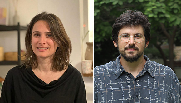
Prof. Michael Gilead (Tel Aviv University) and Dr. Gal Gutman (Ben-Gurion University of the Negev)
The models generated biographies for both Jewish names—such as Ethan Katz, Noah Weiss, and Gabriel Horowitz—and non-Jewish names, including Tyler Johnson, Kyle White, and Dylan Wilson.
Below are excerpts from two example biographies:
The researchers then removed all names and references to religion before asking the AI models to evaluate each character's personality, social status, and psychological traits, allowing them to determine which characteristics had been influenced solely by the assigned name.
The results showed that characters with Jewish names were consistently perceived as more intelligent, more efficient, more assertive, and stronger leaders. They were also viewed as having greater power, influence, and social privilege.
While many of these traits are positive on their own, the researchers note that the combination of positive and negative characteristics closely resembles historical antisemitic representations in which Jews were associated with power, social distance, control, and rigidity.
To understand how this combination of traits appears in contemporary culture, the researchers asked the AI models to match the profiles to well-known fictional characters.
The models repeatedly identified Sherlock Holmes, Dr. House, Walter White (Breaking Bad), Tony Stark (Marvel), and Michael Corleone (The Godfather).
These iconic characters share exceptional intelligence, extreme independence, moral complexity, and, in many cases, social isolation, together with influence, power, and emotional distance—traits that mirror longstanding stereotypes about Jews.
Prof. Gilead explains:
"For most of history, these tropes circulated through pamphlets, caricatures, and rumor. Today they sit, dormant but intact, inside systems that hundreds of millions of people consult every day. The models never say anything explicitly antisemitic, but they may be predisposed to evaluate Jewish individuals in a way that replicates ancient antisemitic tropes."

Iron Man and The Godfather – fictional characters sharing the same stereotypical traits associated with Jewish names
The findings were replicated using additional AI models and validated by hundreds of participants from the United States. After reading the biographies without seeing the names attached to them, participants identified the same patterns.
According to Dr. Gutman:
"Artificial intelligence systems do not express antisemitism in an intentional or conscious sense. Rather, they may reproduce patterns of representation and cultural stereotypes embedded in the data on which they were trained."
She adds:
"Historical biases do not simply disappear; they can persist within the deep structure of the knowledge these models learn, even after alignment and bias-mitigation processes. Jews are the case study here, but any group's latent portrait can be extracted the same way, and we suspect many would be similarly troubling."
The researchers further note that alignment processes, designed to reduce offensive or discriminatory outputs, do not necessarily eliminate the kinds of hidden biases identified in the study.
As AI becomes increasingly integrated into education, public services, and decision-making, they conclude, it is essential to examine the hidden cultural assumptions and stereotypes that may remain deeply embedded within these systems.

Research
A new TAU study finds that while the government's flagship cost-of-living program lowered prices on selected products, it was accompanied by price increases across dozens of other categories.
Researchers from the Coller School of Management at Tel Aviv University examined the Ministry of Economy's flagship "Israel's Basket" initiative and found that although it substantially reduced the prices of the 100 products included in the program, these reductions were accompanied by price increases across many other product categories. According to the researchers, the overall impact on consumers' grocery bills may therefore have been considerably smaller than expected.
The study was conducted by Prof. Itai Ater and Adi Omer of the Coller School of Management at Tel Aviv University, together with Dr. Or Avishay-Rizhi of Pompeu Fabra University in Barcelona. Based on daily price data collected by the Pricez platform from approximately 70 supermarket chains and more than 2,000 stores across Israel, the researchers examined the program's effects during its first seven weeks.
As part of the initiative, Carrefour committed to selling a basket of 100 products for a total price of NIS 1,098, compared with an average price of approximately NIS 1,700 before the program was launched. In return, the Israeli government allocated NIS 50 million to support a nationwide advertising campaign. The initiative operates in 50 Carrefour branches and on the company's online shopping platform for a period of six months.
Analysis of more than 2,000 stores and Pricez data showed that prices of the designated basket products fell by approximately 35% in participating Carrefour branches. Prices also declined by approximately 6% in Carrefour branches that were not participating in the initiative, suggesting that some of the price reductions spilled over to other parts of the chain. On Carrefour's online platform, basket products recorded an average price reduction of approximately 37%.

Carrefour in Israel
The researchers also examined price changes across product categories that were not included in the government's basket.
Among participating Carrefour stores, prices increased in 46 of the 76 product categories examined, with 23 categories showing statistically significant increases. Notable examples included:
The pattern was even more pronounced online, where prices increased in 55 product categories. Among the largest increases were:
The researchers also found price increases of approximately 1.5% to 2% for a range of other popular products that were not included in the government basket.
Outside Carrefour, the program's impact was considerably more limited. When the researchers examined physical stores across Israel's supermarket chains, they found no statistically significant reduction in the prices of the designated basket products. Focusing specifically on the country's major discount chains, they identified an average price reduction of approximately 3%.
Among the retailers:
By contrast, the researchers found no statistically significant price reductions at Shufersal Deal, Osher Ad, or Universe.
The online market showed a similar pattern. Competing retailers reduced the prices of basket products by an average of only 2.3%, with substantial differences between chains:
No significant price reductions were observed at Shufersal Online, the largest online grocery retailer in Israel, or at Mahsanei HaShuk Online.

Does Shopping Near a Carrefour Store Make a Difference?
The researchers also examined whether competing supermarket chains responded differently in areas where Carrefour stores participating in the initiative were located.
Their findings showed no meaningful differences between cities with participating Carrefour branches and those without them. According to the researchers, this suggests that Israel's major supermarket chains set prices primarily at the national level, rather than competing store by store.
In other words, the presence of a nearby Carrefour branch offering discounted basket products did not lead competing supermarkets in the same city to lower their prices.
Prof. Itai Ater concludes:
"There is no doubt that the program succeeded in lowering the prices of the designated basket products in the relevant Carrefour branches and also led to some price reductions among certain competitors. But in economics, there are no miracles and there are no free gifts.
"When you look beyond the 100 products at the center of the campaign, you see that these price reductions were accompanied by increases across dozens of other product categories. As a result, consumers who saved money on the basket products paid more for other items.
"The real test is not the price of a single product, but the total amount consumers pay at the checkout. From that perspective, it is possible that the effort to reduce the cost of the shopping basket ultimately produced the opposite result."

Prof. Itai Ater (Photo: Oded Antman)
Prof. Itai Ater of the Coller School of Management is a leading researcher in the fields of competition policy, industrial organization, and public policy. His research examines the economic mechanisms that shape markets and influence consumers' everyday spending.

Research
New TAU study reveals how tumors reprogram immune cells to support their own growth, opening the door to new treatment strategies.
Researchers at Tel Aviv University’s Gray Faculty of Medical and Health Sciences have uncovered how a natural and essential immune system process can be hijacked to promote cancer progression. In a new study, the research team developed an advanced technology that enabled them to track macrophages, immune system cells, in real time and reveal how they alter their behavior within a cancerous tumor after consuming dead cancer cells. Their findings may pave the way for the development of new treatments targeting the specific macrophages identified in the study, restoring the immune system’s ability to fight the tumor instead of helping it.
The study was led by Dr. Merav Cohen and doctoral students Roi Balaban and Ori Moskowitz of the Gray Faculty of Medical and Health Sciences at Tel Aviv University. It was published in the prestigious journal Science Immunology.
At the heart of the study are macrophages, immune cells whose role is to clear the body of damaged and dead cells. This process is essential for maintaining tissue health and preventing inflammation. However, the researchers discovered that within the environment of a cancerous tumor, this same mechanism can take an unexpected turn: instead of helping the body, it causes immune cells to adopt characteristics that actively support tumor growth.
To understand how this occurs, the researchers developed an innovative method called Effero-seq, which enables the tracking of changes that occur in immune cells after they engulf dead cells. Using this technology, the team found that as the process progresses, the immune cells undergo "reprogramming" and begin activating genes that promote tumor development.
Using a melanoma model, the researchers found that macrophages that had consumed dead cancer cells stimulated the formation of new blood vessels within the tumor. These blood vessels supply the tumor with oxygen and nutrients, enabling it to grow more rapidly. At the same time, these macrophages lost some of their ability to respond to signals that trigger anti-cancer immune activity.
The researchers also analyzed data from patients with uveal melanoma, a form of eye cancer. They found that patients whose tumors contained higher expression of immune cells bearing the genetic signature identified in the study tended to have lower survival rates.
According to Dr. Cohen, the findings provide a new perspective on how cancerous tumors manipulate their surroundings and harness the immune system for their own needs. “The better we understand these mechanisms, the better equipped we will be to develop treatments that block them and restore the immune system’s ability to fight cancer,” she says. “This research points to a new and promising therapeutic target, one that focuses not only on the cancer cells themselves, but also on the processes that enable them to thrive.”

Research
Tel Aviv University researchers identified a rare group of cells with the potential to regenerate sensory hair cells in the inner ear.
A groundbreaking study by a team of researchers from the Gray Faculty of Medical and Health Sciences at Tel Aviv University offers new hope to millions of people suffering from irreversible hearing loss. The researchers have identified a unique biological mechanism that could, in the future, enable the regeneration of sensory hair cells in the inner ear - a process previously thought to be impossible in humans.
The study was conducted under the leadership of Prof. Karen Avraham, Dean of the Gray Faculty of Medical and Health Sciences Drs Sarah and Felix Dumont Chair for Research of Hearing Disorders incumbent. It was spearheaded by Lama Khalaily, a Tel Aviv University doctoral student, in collaboration with Prof. David Sprinzak of TAU’s Wise Faculty of Life Sciences, Shahar Kasirer from Prof. Sprinzak’s laboratory, Dr. Litao Tao of Creighton University in Omaha, and additional researchers. The findings were published in the journal Science Advances.
Hearing loss is often caused by damage to hair cells in the cochlea - cells responsible for detecting sound and converting it into electrical signals transmitted to the brain. Unlike many other species, mammals, including humans, are unable to regenerate these cells once they are damaged, making the loss permanent.
Using live tissue imaging and single-cell multi-omics methods, the researchers focused on supporting cells - cells adjacent to the hair cells that, under normal conditions, cannot regenerate or transform into hair cells. To explore whether and how this limitation could be overcome, the research team inhibited the Notch signaling pathway, a key communication mechanism between cells that is responsible for hair cell differentiation during embryonic development. The team uncovered a rare subset of supporting cells with an unexpected regenerative potential. Rather than responding uniformly, only a distinct group of cells entered a transitional state and began converting into hair cells.
These cells, termed transdifferentiating Deiters’ cells (tDCs), are capable of making the transition from supporting cells to hair cells - a step that is essential for hair cell regeneration. The researchers found that these cells exhibit unique genetic and epigenetic characteristics, enabling them to respond to stimulation and initiate the regeneration process.

Live imaging of the cochlear sensory epithelium: Supporting cells are shown in green and hair cells in red.
The researchers note that a deeper understanding of the mechanisms that allow certain cells to regenerate may pave the way for the development of innovative treatments that activate this regenerative ability in additional cells. Future approaches may involve a combination of genetic and epigenetic interventions designed to bypass existing biological barriers. According to the research team, this represents a significant step toward the development of regenerative treatments for hearing loss - a field in which no restorative medical solutions currently exist, only assistive measures such as hearing aids and cochlear implants and limited gene therapy.
Prof. Karen Avraham concludes: “Our study shows that even in tissues long considered incapable of regeneration, such as the cochlea of the inner ear, there is in fact a hidden regenerative capacity, though it is very limited and appears only in a rare subpopulation of cells. The major challenge now is to understand how this ability can be expanded and activated in additional cells. If we succeed in doing this, we may lay the foundation for the development of innovative biological treatments that restore hearing, rather than merely compensate for its loss. This is a first but significant step toward a deeper understanding of regeneration in the auditory system and in neural systems in general.”
The study was supported by a Breakthrough Research grant from the Israel Science Foundation, the Ernest and Bonnie Beutler Research Program of Excellence in Genomic Medicine, and the Sagol Center for Regenerative Medicine at Tel Aviv University.


")
