TAU researchers have developed a targeted method of delivering locked nucleic acids (LNAs) using lipid nanoparticles, achieving therapeutic effect against inflammatory bowel disease in preclinical models — without side effects.


Research
TAU researchers have developed a targeted method of delivering locked nucleic acids (LNAs) using lipid nanoparticles, achieving therapeutic effect against inflammatory bowel disease in preclinical models — without side effects.
Researchers at Tel Aviv University have developed a new approach for using locked nucleic acids (LNAs) – a particularly stable type of RNA – to treat inflammatory bowel diseases such as Crohn’s disease and ulcerative colitis. The researchers encapsulated selected LNA molecules, which silence a key gene in colitis, within lipid (fat) nanoparticles that serve as targeted drug carriers and injected the nanoparticles into colitis-model mice. The findings indicated improvement in all markers of systemic inflammation, with no side effects. According to the researchers, this innovative method may also be suitable for a wide range of other diseases – including rare genetic disorders, vascular and heart diseases, and neurological diseases such as Parkinson’s and Huntington’s.
The study was conducted by the group of Prof. Dan Peer, a pioneer in the use of RNA molecules for therapy and vaccines, world expert in nanomedicine, and a senior faculty member at TAU's Shmunis School of Biomedicine and Cancer Research, Department of Materials Sciences and Engineering at the Fleischman Faculty of Engineering, Jan Koum Center for Nanoscience and Nanotechnology, and Cancer Biology Research Center. The group, led by Neubauer doctoral student Shahd Qassem together with Dr. Gonna Somu Naidu, a postdoctoral fellow who collaborated with researchers from F. Hoffman La-Roche (Roche) pharmaceutical company in Switzerland. The article was published in Nature Communications.
Prof. Peer explains: “Our study focused on unique RNA molecules called LNA. Unlike most RNA molecules, LNA molecules are very stable and do not break down easily. Consequently, until about 10 years ago, they were thought to have great potential as genetic drugs. However, experiments in laboratory animals, as well as clinical trials in humans (in chronic liver inflammation), showed that very large amounts of LNA are needed to achieve therapeutic efficacy. Moreover, administered by injection as a free drug, this high dosage proved very costly and caused severe side effects when spreading throughout the body. As a result, the effort to develop LNA-based drugs was abandoned. In our study we sought to test a new, better targeted and more effective approach.”

Prof. Dan Peer
The researchers used a method previously developed at Prof. Peer’s lab for other RNA molecules (such as siRNA, mRNA, circRNA), now applying it to LNA: they encapsulated the molecules in lipid nanoparticles (LNPs) that serve as targeted drug carriers, delivering their therapeutic payload directly to the relevant organ in the body. Specifically, they chose an LNA molecule known to silence the TNFα gene, which plays a significant role in inflammatory bowel diseases. Screening a lipid library developed in Prof. Peer’s lab over the past 13 years, they identified the most suitable lipid molecules and encapsulated the LNA molecules in them. The resulted LNPs were injected into mice in a model of chronic bowel diseases such as colitis.
The findings were highly encouraging: the dosage required to achieve the desired therapeutic effect was 30 times lower compared to past studies – in which LNA molecules were administered as a free drug without lipid encapsulation. At the current dosage, delivered precisely to the correct site, the drug proved highly effective in treating the disease, without causing any side effects.
Prof. Peer: “Our study paves the way to developing new LNA-based drugs for inflammatory bowel diseases, as well as a wide range of other diseases – including rare genetic disorders, vascular and heart diseases, and neurological diseases such as Parkinson’s and Huntington’s. So far, we have demonstrated that the new method is effective in chronic bowel inflammation in mice. We hope to proceed to clinical trials in humans in the near future.”

Research
A new TAU–Ichilov study shows that tracking eye movements can assess memory more accurately than verbal reports, with potential use for infants, Alzheimer’s patients, and brain injury victims.
Researchers from Tel Aviv University and Tel Aviv Sourasky Medical Center (“Ichilov”) have measured subjects' memory without asking whether they remembered something or not - -simply by tracking their eye movements as they watched animation videos. The study demonstrated that people actually remember more than they report. Moreover, this method can be used to measure memory in subjects who cannot speak— including infants, patients with brain injuries, and even animals.
The groundbreaking study was led by Dr. Flavio Jean Schmidig, Daniel Yamin, Dr. Omer Sharon, and Prof. Yuval Nir from the Sagol School of Neuroscience, the Gray Faculty of Medical and Health Sciences, and the Fleischman Faculty of Engineering at Tel Aviv University, as well as the Sagol Brain Institute at the Tel Aviv Sourasky Medical Center (“Ichilov”). The paper was published in Communications Psychology.
"Memory is usually tested through direct questioning, with subjects verbally reporting whether they remember a certain event," explains Dr. Flavio Schmidig, currently completing his postdoctoral research in Prof. Yuval Nir’s lab at TAU. "For example, a subject might be shown a picture and asked if they remember having seen it before. However, this type of testing cannot be performed on animals, infants, patients with advanced Alzheimer’s, or people with head injuries who cannot speak. In this study we wanted to test memory in a more natural way, without asking people to remember."
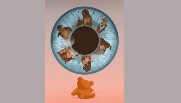
"Gaze Memory" Illustration by Ana Yael
In the study, 145 healthy subjects watched specially created animation videos that included a surprising event - for example, a mouse suddenly jumping out of the corner of the frame. Tracking the subjects' eye movements across two separate viewings of the same films, the researchers found that during the second viewing, subjects shifted their gaze toward the area where the surprising event was about to occur. A comparison of eye movement data with verbal memory reports indicated that gaze direction was in fact a more accurate measure. In some cases, subjects said they did not remember the mouse, yet their gaze indicated that they did.
"The study proves that tracking eye movements can be an excellent alternative to verbal questions such as 'Do you remember this?'," says Daniel Yamin. "In a series of experiments, we demonstrated that gaze direction is a very sensitive gage of memory. Even when subjects said they didn’t remember, their gaze direction showed they did. This means that sometimes people remember, but can't say that they remember. By using AI machine learning techniques, it is possible to infer automatically, from just a few seconds of eye tracking, whether someone has seen a video before and formed a memory of it."
"When I ask you if you remember," adds Dr. Sharon, "you might give any of several answers: yes, no, not sure, etc. But when you look to the left of the frame due to a vague memory that something is about to happen there, finer nuances can be discerned. Now we have a tool for testing to what extent memory is present. Our new method is also more natural than traditional memory tests."
"The results of this study are especially relevant when verbal reports on memory cannot be obtained," adds Prof. Yuval Nir, the study's supervisor. "We believe that in the future this new method may be used for measuring memory functions in infants, Alzheimer's patients, and people with brain injury whose speech ability has been impaired. Gaze direction can be simply detected by the camera of a laptop or smartphone as the subject views a video - with no need for large, sophisticated equipment. The method has the potential for identifying memories even in situations that have so far been out of reach for us as scientists and clinicians."
: Roni Hahn & Prof. Karen Avraham.")
Research
TAU researchers created a gene therapy that protects against hearing and balance impairments caused by inner ear dysfunction.
Scientists from the Gray Faculty of Medical & Health Sciences at Tel Aviv University introduced an innovative gene therapy method to treat impairments in hearing and balance caused by inner ear dysfunction. According to the researchers, “This treatment constitutes an improvement over existing strategies, demonstrating enhanced efficiency and holds promise for treating a wide range of mutations that cause hearing loss.”
The study was led by Prof. Karen Avraham, Dean of the Gray Faculty of Medical & Health Sciences, and Roni Hahn, a PhD student from the Department of Human Molecular Genetics and Biochemistry. The study was conducted in collaboration with Prof. Jeffrey Holt and Dr. Gwenaëlle Géléoc from Boston Children's Hospital and Harvard Medical School and was supported by the US-Israel Binational Science Foundation (BSF), the National Institutes of Health/NIDCD and the Israel Science Foundation Breakthrough Research Program. The study was featured on the cover of the journal EMBO Molecular Medicine.
Prof. Avraham explains: “The inner ear consists of two highly coordinated systems: the auditory system, which detects, processes, and transmits sound signals to the brain, and the vestibular system, which enables spatial orientation and balance. A wide range of genetic variants in DNA can affect the function of these systems, leading to sensorineural hearing loss and balance problems. Indeed, hearing loss is the most common sensory impairment worldwide, with over half of congenital cases caused by genetic factors. In this study, we aimed to investigate an effective gene therapy for these cases using an approach that has not been applied in this context before.”
Roni Hahn: “Gene therapy has emerged as a powerful therapeutic approach in recent years and is now being applied to a range of genetic disorders, including spinal muscular atrophy (SMA) and Leber congenital amaurosis (LCA), as well as in cancer immunotherapy approaches such as CAR T-cell therapy. One of the treatment strategies includes the use of engineered viral vectors, in which the native DNA is replaced with a functional sequence of the target gene. These vectors utilize the virus's natural ability to enter cells to deliver the correct gene sequence, thereby restoring normal function. Many gene therapies utilize adeno-associated viruses (AAVs) to introduce therapeutic genetic material into target cells, and AAV-based gene therapy for hearing loss is currently in clinical trials, showing promising early results.
In this study, the researchers investigated a mutation in the CLIC5 gene, which is essential for maintaining the stability and function of hair cells in the auditory and vestibular systems. Deficiency of this gene causes progressive degeneration of hair cells, initially leading to hearing loss and later resulting in balance problems.
The researchers utilized an advanced, structurally optimized version of the AAV vector, the self-complementary AAV (scAAV). They found that this vector achieved faster and more efficient transduction of hair cells compared to traditional AAV methods, requiring a lower dose to achieve a similar therapeutic effect. In treated animal models, this approach prevented hair cell degeneration and preserved normal hearing and balance.
In summary, Prof. Avraham states: “In this study, we applied an innovative treatment approach for genetic hearing loss and found that it improves therapeutic effectiveness while also addressing combined impairments in hearing and balance. We anticipate that these findings will pave the way for developing gene therapies to treat a wide range of genetically caused hearing disorders."

Research
With a new method called photonic origami, researchers can bend ultra-thin glass sheets into complex, ultra-smooth structures directly on a chip — a step toward new optical devices for data processing, sensing, and experimental physics.
Researchers explain that traditional 3D printers produce rough structures that lack the smoothness and optical uniformity required for high-performance optics. To overcome this limitation, the TAU team devised a laser-induced technique inspired by nature — similar to how a pinecone’s scales bend outward to release seeds. By triggering precise bends in ultra-thin glass sheets, the method creates highly transparent, ultra-smooth 3D microphotonic devices suitable for a wide range of applications.
“Existing 3D printers produce rough 3D structures that aren’t optically uniform and thus can’t be used for high-performance optics,” said research team leader Prof. Tal Carmon from the School of Electrical Engineering, Fleischman Faculty of Engineering, at Tel Aviv University in Israel. “Mimicking the way a pinecone’s scales bend outward to release seeds, our laser-induced technique triggers precise bending in ultra-thin glass sheets and can be used to create highly transparent, ultra-smooth 3D microphotonic devices for a variety of applications.”
In Optica, Optica Publishing Group’s journal for high-impact research, the researchers reported that the new laser-induced folding method can create 3-mm-long structures just 0.5 microns thick — about 1/200th the width of a human hair — setting a record length-to-thickness ratio of 3D structures. They also created helix shapes as well as concave and convex mirrors with surfaces so smooth — less than a nanometer of variation — that light reflects off them without distortion.
“Similar to how large 3D printers can fabricate almost any household item, photonic origami could enable a variety of tiny optical devices,” said Carmon. “For example, it can be used to generate micro-zoom lenses that could replace the five separate cameras used in most smartphones or to fabricate microphotonic components that use light instead of electricity — helping drive the shift toward faster, more efficient alternatives to traditional electronics in our computers.”
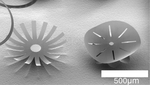
Structures made with photonic origami
The new method was discovered by chance when Carmon asked graduate student Manya Malhotra to pinpoint where an invisible laser was hitting the glass by increasing the power until the spot glowed. Instead of glowing, the glass folded — revealing a simple and unexpected way to achieve glass folding. Malhotra then became the pioneering expert in photonic origami.
The glass folds because, as one side is heated with a laser, the glass liquifies and surface tension becomes stronger than gravity. As the surface tension increases, the glass is pulled into a fold precisely where the laser hits.
To apply this discovery, lab engineer Ronen Ben Daniel fabricated a thin layer of silica glass on a silicon chip and then shaped it into the required two-dimensional form. Before bending the glass, the researchers used etching to undercut the silicon beneath the glass sheet while leaving a small support region to hold it in place. Using CO2 laser pulses, they showed that thin glass sheets on a silicon chip could be folded in less than a millisecond, with a speed of 2 m/s and acceleration exceeding 2000 m/s2.
“It was exciting to see the folding silica under the microscope,” said Carmon. “The level of control we had over 3D microphotonic architecture came as a pleasant surprise — especially given that it was achieved with a simple setup involving just a single laser beam focused on the desired fold.”

Folding glass bar
Using the new photonic origami approach, the researchers were able to bend sheets of glass up to 10 microns thick into shapes ranging from a 90-degree knee to helices. They were able to do this with fine control, down to 0.1 microradians.
They also used the new approach to create an extremely lightweight and precise table structure containing a concave cavity mirror, a type of mirror that focuses light. This structure was inspired by a theoretical paper by P.K. Lam from the Australian National University that proposed exploring potential deviations from Newtonian gravity at very small scales using optically levitated cavity mirrors that might be possible to fabricate using photonic origami.
To make the tiny table light enough, the researchers began with a glass sheet just 1/20 the thickness of a human hair (5 microns). They patterned the sheet much like a child’s foldable paper table toy and used their photonic origami technique to fold it into a 3D table after fabricating a concave mirror at the base of the table.
According to the researchers, this ultra-light, compact table could, in principle, be optically levitated and used to explore possible deviations from Newtonian gravity. These types of experiments could provide insights into astronomical mysteries associated with dark matter —the only area in physics where experimental observations consistently defy current theoretical predictions.
“High-performance, 3D microphotonics had not been previously demonstrated,” said Carmon. “This new technique brings silica photonics — using glass to guide and control light — into the third dimension, opening up entirely new possibilities for high-performance, integrated optical devices.”
Optica is an open-access journal dedicated to the rapid dissemination of high-impact peer-reviewed research across the entire spectrum of optics and photonics. Published monthly by Optica Publishing Group, the Journal provides a forum for pioneering research to be swiftly accessed by the international community, whether that research is theoretical or experimental, fundamental or applied. Optica maintains a distinguished editorial board of more than 60 associate editors from around the world and is overseen by Editor-in-Chief Prem Kumar, Northwestern University, USA. For more information, visit Optica.
Optica Publishing Group is a division of the society, Optica, Advancing Optics and Photonics Worldwide. It publishes the largest collection of peer-reviewed and most-cited content in optics and photonics, including 18 prestigious journals, the society’s flagship member magazine, and papers and videos from more than 835 conferences. With over 400,000 journal articles, conference papers and videos to search, discover and access, our publications portfolio represents the full range of research in the field from around the globe.

Research
TAU researchers begin to prepare for implantation of engineered spinal cord tissues in humans — a new development that brings fresh hope to paralyzed patients.
What if we could restore the ability to walk to people paralyzed by injury or illness?
This vision is now moving closer to reality. Three years ago, Tel Aviv University researchers succeeded in engineering a human spinal cord in the lab for the first time. Since then, progress has been rapid, with animal trials showing unprecedented success. Now, for the first time, the technology is set to be tested in human patients.
Prof. Tal Dvir, of TAU’s Sagol Center for Regenerative Biotechnology, head of TAU’s Jan Koum Center for Nanoscience and Nanotechnology, and Chief Scientist of the biotech company Matricelf, explains:
“The spinal cord is made up of nerve cells that transmit electrical signals from the brain to every part of the body. When the spinal cord is torn due to trauma — from a car accident, a fall, or a battlefield injury — this chain is broken. Think of it like an electrical cable that’s been cut: if the two parts don’t touch, the electrical signal can’t pass. The cable won’t carry electricity, and in the same way, the person can’t transmit the signal beyond the site of the injury.”
This is one of the few injuries in the human body with no natural ability to regenerate. “Neurons are cells that do not divide and do not renew themselves. They are not like skin cells, which can repair themselves after injury. They are more similar to heart cells: once damage occurs, the body cannot restore them,” notes Prof. Dvir.
To overcome this challenge, the TAU researchers developed a fully personalized process. Blood cells are taken from the patient and reprogrammed through genetic engineering to behave like embryonic stem cells, capable of becoming any type of cell in the body.
Meanwhile, fat tissue from the same patient is used to extract substances such as collagen and sugars. These are used to produce a unique hydrogel. “The beauty of this gel is that it’s also personalized, just like the cells. We take the cells that we’ve reprogrammed into embryonic-like stem cells, place them inside the gel, and mimic the embryonic development of the spinal cord,” says Prof. Dvir.
The result is a complete three-dimensional implant. “At the end of the process, we don’t just turn the cells into motor neurons — because cells alone won’t help us — but into three-dimensional tissue: neuronal networks of the spinal cord. After about a month, we obtain a 3D implant with many neurons that transmit electrical signals. These 3D tissues are then implanted into the damaged area.”

Visualization of the next stage of the research - human spinal cord implants for treating paralysis (Photo: Sagol Center for Regenerative Biotechnology)
The researchers first tested the implant in lab animals. “We showed that we can treat animals with chronic injuries. Not animals that were injured just recently, but those we allowed enough time to pass — like a person more than a year after an injury. More than 80% of the animals regained full walking ability,” Prof. Dvir explains.
Encouraged by these results, the team submitted the findings to Israel’s Ministry of Health. “About six months ago we received preliminary approval to begin compassionate-use trials with eight patients. We decided, of course, that the first patient would be Israeli. This is undoubtedly a matter of national pride. The technology was developed here in Israel, at Tel Aviv University and at Matricelf, and from the very beginning it was clear to us that the first-ever surgery would be performed in Israel, with an Israeli patient.” he says.
The first implant in a human patient is expected within about a year. For the initial trials, the team will focus on patients whose paralysis is relatively recent — within about a year of injury. “Once we prove that the treatment works — everything is open, and we’ll be able to treat any injury,” says Prof. Dvir.
Behind the initiative are key figures from both academia and industry. Prof. Dvir founded Matricelf in 2019 together with Dr. Alon Sinai, based on the revolutionary organ engineering technology developed at TAU under a licensing agreement through Ramot, the University’s technology transfer company. The company’s CEO is Gil Hakim, while the scientific development is led by Dr. Tamar Harel-Adar and her team.
“They managed to get us to the stage of regulatory approvals so quickly — and that’s amazing,” says Prof. Dvir.
Gil Hakim, CEO of Matricelf , concludes: "This milestone marks the shift from pioneering research to patient treatment. For the first time, we are translating years of successful preclinical work into a procedure for people living with paralysis. Our approach, using each patient’s own cells to engineer a new spinal cord, eliminates key safety risks and positions Matricelf at the forefront of regenerative medicine. If successful, this therapy has the potential to define a new standard of care in spinal cord repair, addressing a multi-billion-dollar market with no effective solutions today. This first procedure is more than a scientific breakthrough, it is a value-inflection point for Matricelf and a step toward transforming an area of medicine long considered untreatable. We are proud that Israel is leading this global effort and are fully committed to bringing this innovation to patients worldwide.”

Research
A TAU-led study finds that seeing an image more than once, real or AI-generated, makes us more likely to believe it’s real.
The study— published in the Journal of Experimental Psychology: Learning, Memory, and Cognition, a prestigious scientific journal of the American Psychological Association (APA) — was led by Guy Grinfeld, a doctoral student at the School of Psychological Sciences, Gershon H. Gordon Faculty of Social Sciences at Tel Aviv University, in collaboration with researchers from Germany, Belgium, and Spain.
The researchers found that repeated images are more likely to be believed as representing a real person, location, or event than images seen for the first time — even when those images were entirely AI-generated. “The study is based on a well-known psychological phenomenon called the ‘mere exposure effect,’ which suggests that information that we encounter repeatedly is perceived as more credible,” Grinfeld explains. “In our research, we sought to examine whether this effect also applies in the visual domain — specifically with images created using artificial intelligence algorithms.
This is the first study to demonstrate the mere exposure effect for images; until now, it had only been demonstrated for text. The findings raise concerns about the spread of false visual information on social media and its influence on public perception. As we like to summarize it, if until now the proverb went, ‘A lie told often enough becomes the truth,’ our study shows that ‘An image seen often enough becomes reality.’”
In the experiment, participants were shown a series of images, both real photographs and AI-generated visuals. Later, they saw some of the same images again along with images they had not seen before, and were asked to judge whether each depicted a real object or event. The result was clear: images that participants had seen before were rated as more credible than new images — regardless of whether they were real or fake.
Surprisingly, the repetition effect was even stronger among the skeptical participants—those who generally rated images as less credible. This suggests that people who tend to be cautious might rely more heavily on repetition as an indicator of truth.
“In the era of social networks and digital media, we are constantly and involuntarily exposed to visual information,” says Grinfeld. “Whereas in the past, it was easy to lie with words, today, AI tools make it just as easy to ‘lie’ with images. Our new study reveals a troubling mechanism: people attribute higher credibility to visual information that is repeated, regardless of its veracity. This creates a dangerous combination: repeated exposure to false information can make it seem credible, simply through repetition.
“The findings raise profound questions about how we process information, especially in an age of visual overload in social and news media. They also highlight the central challenge of our time: preserving truth and critical thinking in a world of dynamic, easily manipulated, and hard-to-discern visual content.”
Guy Grinfeld, lead researcher of the study
: Anish Dsilva & Prof. Ariel Munitz")
Research
TAU researchers found that blocking the protein TSLP may prevent a painful, food allergy–driven disease on the rise worldwide.
A new study from the Gray Faculty of Medical and Health Sciences at Tel Aviv University may mark a breakthrough in the treatment of Eosinophilic Esophagitis (EoE) — a chronic inflammatory disease of the esophagus caused by food allergies. EoE leads to difficulty swallowing, chest and abdominal pain, and even growth delays in children. Its prevalence has been steadily increasing over the past decade in Israel and the Western world.
In this study, researchers identified the protein TSLP as a trigger for the disease’s development, and found that neutralizing it may significantly ease symptoms.
The study, led by Prof. Ariel Munitz and doctoral student Anish Dsilva from the Gray Faculty of Medical and Health Sciences, was conducted in collaboration with Dr. Chen Varol of Ichilov Hospital, Prof. Marc Rothenberg of Cincinnati Children’s Hospital, and the pharmaceutical company AstraZeneca. It was supported by grants from the Israel Science Foundation, the US-Israel Binational Science Foundation, and the Azrieli Foundation Canada–Israel. The article was published in Allergy, the leading journal in clinical immunology.
Prof. Munitz explains: “Eosinophilic Esophagitis, or EoE, is a type of food allergy. It is a chronic inflammation of the esophagus caused by an abnormal immune response to food — mainly milk, eggs, wheat, nuts, fish, and more. The disease is characterized by an accumulation of eosinophils, a type of white blood cell that is not typically present in a healthy esophagus. EoE is often associated with other allergic conditions such as asthma and atopic dermatitis. It causes difficulty swallowing, food getting stuck in the esophagus, chest and abdominal pain, and growth delays in children. Current treatments require restrictive diets, and in severe cases, patients rely on essential amino acid formulas. Over the past decade, there has been a concerning rise in the prevalence of EoE worldwide, including in Israel. We are studying the disease in depth to understand the involvement of various immune system components in its progression. These components may serve as targets for future treatment for this disease, and for other allergic disorders as well.”
A previous study from Prof. Munitz’s lab, also published in Allergy, presented an experimental model that closely mimics the course and symptoms of EoE in humans. As a direct continuation of that study, the researchers now focused on a specific aspect of the disease, aiming to understand the role of epithelial cells. Prof. Munitz elaborates: “Epithelial cells form a protective outer layer that prevents foreign bodies from entering organs, including the digestive and respiratory systems. In allergic conditions, epithelial cells release various substances in response to encountering an allergen, and these substances trigger the chain of events that initiate the inflammatory process we experience as an allergy attack.”
The researchers found that epithelial cells in the esophagus of the EoE experimental model secreted high levels of two proteins: IL-33 and TSLP. They also discovered that the esophageal tissue contained immune cells with receptors for both proteins, indicating that these are active proteins capable of initiating the disease.
They then examined whether each protein had a distinct role or acted together. Using genetic engineering, they created models lacking one of the proteins.
The results were clear: removing IL-33 did not change the disease course, but removing TSLP led to a dramatic improvement — in some cases preventing the disease entirely. Similarly, neutralizing TSLP with an antibody caused a significant reduction in symptoms. Sequencing and bioinformatic analyses confirmed that TSLP acts as a master regulator of EoE, making it a promising therapeutic target.
Prof. Munitz concludes: “In this study, we found that the TSLP protein is a central player in EoE — a disease that causes significant suffering and is becoming increasingly prevalent worldwide. We know that pharmaceutical companies are currently developing a variety of antibodies targeting disease-causing proteins, under the broad category of biological therapies, including antibodies against TSLP. We believe these antibodies could serve as an effective treatment for EoE.”