


An international team led by TAU’s Prof. Oded Rechavi is recreating century-old experiments to explore how traits can be inherited beyond genetics.


Research
An international team led by TAU’s Prof. Oded Rechavi is recreating century-old experiments to explore how traits can be inherited beyond genetics.
In 1902, three prominent Jewish biologists established the Biologische Versuchsanstalt (BVA) in what was then Austro-Hungarian Vienna. Now, an international team led by Tel Aviv University’s Prof. Oded Rechavi has been awarded a $1.2 million grant by the prestigious Human Frontier Science Program (HFSP) to continue their work . HFSP is known for its highly competitive selection process, approving only 4% of proposals submitted each year, and indeed the team’s project is truly exceptional, both scientifically and historically.
“We propose a unique study, combining history and cutting-edge biology, focused on the BVA - one of the most groundbreaking institutes of the early 20th century,” says Prof. Rechavi, of the School of Biochemistry, Neurobiology, and Biophysics at the Wise Faculty of Life Sciences, Tel Aviv University. The institute was notable for conducting long-term experiments in live animals, a new concept in biological study of those days, and its founders, led by Hans Leo Przibram, emphasized the importance of biology as an empirical and quantitative science on the one hand, and of studying animals in habitats as natural as possible on the other. Przibram led and supported very long experiments in hundreds of species of animals, many of which were never used in research later on or even successfully raised in captivity. The BVA was also innovative with the implementation of advanced methods for climate control, allowing researchers to carefully study the influence of the environment on biology.
“The BVA gained notoriety through Paul Kammerer, who claimed that environmental factors influenced inheritance and was later accused of fraud. However, other respected researchers at the BVA also studied the inheritance of acquired traits—without disproof. Tragically, the scandal and the Nazi persecution of the institute’s Jewish members led to its collapse. As modern genetics emerged, the entire concept of acquired trait inheritance was set aside —until recent discoveries in epigenetics brought it back into scientific discourse,” adds Prof. Rechavi.
For nearly a century, the idea of inheriting acquired traits was considered scientific heresy. But in the past 15 years, research in epigenetic inheritance has breathed new life into this controversial topic. Prof. Rechavi identified a molecular mechanism enabling the transgenerational inheritance of acquired traits in the highly useful model organism, the C. elegans nematode, via small RNA molecules. Now, the next challenge is to demonstrate that similar mechanisms exist across other species - potentially reshaping our understanding of evolution. And this is where the BVA’s historical work becomes newly relevant.
“The papers published by BVA researchers made headlines but were largely ignored because, for a long time, few believed in non-genetic inheritance,” Prof. Rechavi explains. “The question is: can we replicate their experiments using modern tools and knowledge? For example, one of BVA director Hans Przibram’s most promising studies involved growing rats to in warm climate over generations to observe whether the environment can affect their offspring body and tail size. We plan to recreate this experiment as one of our first steps. Today’s improved temperature control systems and the ability to account for genetic variation could allow our team to isolate true epigenetic effects from purely genetic one, potentially validating theories that were ahead of their time more than a century ago.”
International partners
Starting this December, Prof. Oded Rechavi will lead the historical research and assessment of the rich scientific legacy of the BVA, with the support of Prof. Gerd Müller, an expert in the study of the relationship between evolution and development and the editor of a recently published book about the BVA, studying the Viennese sociocultural context at the time of the BVA's founding.
Prof. Katharina Gapp, an expert in the study of environmentally induced traits, their epigenetic underpinnings and inheritance in rodents, will lead the reproduction of Przibram's 1925 rat experiments in rodents of genetically identical backgrounds housed in standardized and temperature-controlled cages and complement these observations with molecular studies on small RNA and the mechanistic underpinnings, aided by the expertise in the Rechavi lab.
Prof. Miguel Vences, an expert in the study of amphibian phylogeny and systematics and patterns and processes of species formation, will lead the reproduction of the salamander experiments conducted in the BVA with the goal of identifying if any of these studies indeed succeeded in demonstrating the inheritance of such environmentally triggered changes.
According to Kammerer, his "experimentum crucis" describing acquired traits inheritance was with a sea squirt (ascidian) called Ciona intestinalis, allegedly demonstrating a transgenerational effect of siphon elongation following amputations. Unfortunately, Kammerer never published his study design, and multiple attempts to reproduce it failed to identify transgenerational effects. Prof. Yasunori Sasakura, a world leader in the study of ascidians as models for developmental genetics and evolution, who was the first to make knockout strains of Ciona intestinalis, will lead the search for molecular mechanisms of epigenetic inheritance in the model organism. C. intestinalis is an organism with large phenotypical diversity in different environmental conditions. Identification of such epigenetic inheritance mechanisms in the ascidian could provide an indication to the validity of the experiments conducted at the BVA without reproducing them precisely.

Research
A lab-grown kidney model is giving researchers a rare view into human development, birth defects, and potential new therapies.
For the first time researchers from both Tel Aviv University and Sheba Medical Center have grown human kidney organoids ,a synthetic 3D organ culture, from tissue stem cells in the laboratory mirroring human fetal kidney development.
The kidney grew and developed over months, allowing researchers to see the development of the organ in real time, isolate genes that lead to birth defects, develop new treatments in the field of regenerative medicine, and test the toxicity of drugs during pregnancy on fetal kidneys.
The current model has matured and stayed stable for over half a year. Previous kidney organoids mimicking development broke down within four weeks. This allows long-term research and medical testing on kidney models.
It is also the purest kidney organoid ever developed, with no cross contamination from stem cell development. Previous models using pluripotent stem cells would develop other cellular structures due to the unstable nature of the stem cells. The new organoid only expresses kidney cells, allowing for clear cause-and-effect experiments.
The groundbreaking study was led by Prof. Benjamin Dekel, Director of the Sagol Center for Regenerative Medicine at Tel Aviv University and Director of the Pediatric Nephrology Unit and the Stem Cell Research Institute at the Safra Children's Hospital at Sheba Medical Center. Also participating in the study were doctoral student Dr. Michael Namestannikov, a graduate of the Physician-Researcher track at the Gray Faculty of Medical and Health Sciences at Tel Aviv University, and Dr. Osnat Cohen-Sontag, a research associate at Sheba Medical Center, as part of Prof. Dekel's research group. The study was published in the prestigious medical publication The EMBO Journal.
"Life begins with pluripotent stem cells, which can differentiate into any cell in the body," explains Prof. Dekel. "In the past, they were able to grow organoids - 3D organ-like cultures - by producing such general stem cells and sorting them into kidneys, but after about a month the kidney in culture died, and the process had to be started again. About a decade ago, my research group was able to isolate for the first time the human kidney tissue stem cells that are responsible for the growth of the developing organ. Now we have succeeded for the first time in growing a human kidney in the form of an organoid from the specific stem cells of the kidney, and this in parallel with the maturation process in the uterus that occurs until the 34th week of pregnancy."

Human fetal kidney cells
Researchers grow organoids in laboratory conditions to study organs in ways that are not possible in humans, but organoids derived from pluripotent stem cells often contain unwanted cells unrelated to the organ being studied that contaminate experiment data. Prof. Dekel’s organoid grew from kidney tissue stem cells in a "clean" manner, since these stem cells differentiate exclusively into kidney tissue. These cells developed into different types of kidney cells, and over half a year formed different tissues of the kidney, such as blood filter cells and kidney and urinary ducts, a process known as tubulogenesis.
"Growing the fetal kidney structures can shed new light on biological processes in general, and in particular on processes that lead to kidney diseases," says Prof. Dekel. "And indeed, when we selectively blocked a certain signaling pathways [in the organoid], we saw how it lead to a birth defect. We are actually seeing live how a developmental problem leads to kidney diseases that are seen in the clinic, which will enable the development of innovative treatments."
The implications that go far beyond research. "The fact that we can grow kidney tissue stem cells outside the body over time opens the door to regenerative medicine, that is, transplanting kidney tissue grown in the laboratory – inside the body or alternatively harnessing signals the organoid secrets for repair and rejuvenation of a damaged kidney ,” said Prof. Dekel. “We now have an essentially inexhaustible source of different kidney cells, and a better understanding of their different roles in kidney development and function.”
Breakthroughs like this represent Israel’s unique place in the world, says Prof. Dror Harats, Chairman of the Sheba Research Authority. "In recent years, we have witnessed attempts to distance Israel from international centers of influence, and scientific successes of this kind are a reminder that our contribution to medical and scientific research is significant and unquestionable."
: Prof. Moran Rubinstein, Prof. Karen Avraham, and Prof. Aviva Fattal-Valevski. Front row (right to left): Daniel Gelber, Mor Yam, Shir Koren.")
Research
A new model developed at TAU following a family's request is helping researchers study a rare brain disorder known to affect only 40 people worldwide.
When the parents of an 8-year-old Israeli boy reached out to Tel Aviv University, a research team at the Gray Faculty of Medical and Health Sciences stepped up. Their mission: to find answers for a devastating genetic condition with no known cure. The result is a breakthrough mouse model that mimics the disease with striking accuracy — and may pave the way for life-saving treatments.
The study was led by Prof. Moran Rubinstein and Prof. Karen Avraham, Dean of the Faculty. Other participants included students Mor Yam, Julan Nasir, Daniel Gelber, Shir Kavin, Roni Gal, Mor Ovadia, Mor Bordinik-Cohen, and Eden Peled — all from the Gray Faculty of Medical and Health Sciences at Tel Aviv University or the Sagol School of Neuroscience — as well as Dr. Moran Heusman-Kedem and Prof. Aviva Fattal-Valevski from the Pediatric Neurology Institute at Dana-Dwek Children’s Hospital, Tel Aviv Medical Center, and Prof. Christopher McKinnon and Prof. Wayne Frankel from Columbia University in the United States.
Prof. Avraham explains: “We were approached by the parents of an Israeli child named Adam, now 8 years old, who is one of approximately 40 people worldwide suffering from an extremely rare genetic disease. It’s a mutation in a gene called GRIN2D, which causes developmental epilepsy, severe delays in motor and cognitive development, and sometimes even premature death.”
Eden Maimon Benet, Adam’s mother, adds: “At Tel Aviv University, we met a remarkable all-women team that took on the mission: to find a cure for our son. I believe the fact that they got to know Adam and our family personally only deepened their dedication and commitment. When Adam was two years old, we embarked on this long journey together — and today, we can already see real light at the end of the tunnel.”
In the first stage, the researchers aimed to better understand the disease’s characteristics. To do so, they created a mouse model with a mutation similar to that found in human patients. However, due to the severity of the disease, most of the mice did not survive their first weeks of life — before any meaningful research observations could be made. This led the team to conclude that while the model mimics the human disease, it poses a major challenge: too few mice could be generated for scientific study.
To overcome this, they used genetic engineering tools to create a strain of mice that carry the mutation but do not develop symptoms. These serve as carriers, with half of the offspring born healthy and the other half born with the disease. The affected mice exhibited symptoms similar to those seen in children with the disease. Most lived only a few weeks, and only a few survived up to three months. The researchers observed their behaviour and development at four key stages: at two weeks old (infancy), three weeks (when mice transition to solid food — roughly equivalent to a one-year-old child), four weeks (roughly age six in children), and five weeks (the onset of sexual maturity).
“Because the disease is so rare, we don’t yet fully understand how it progresses with age,” Prof. Rubinstein says. “The mouse model helped us characterize symptoms at various stages. The tests we conducted revealed interesting findings: neurological symptoms — including epilepsy, hyperactivity, and severe motor impairments — appeared as early as infancy. Cognitive impairments, on the other hand, showed up later and worsened gradually. In addition, their lifespan was short — most of the affected mice did not survive to sexual maturity.”
In a follow-up experiment, the researchers monitored communication between neurons in the brains of the model mice, focusing on the cerebellum — the brain region responsible for motor control. The tests showed that by just two weeks of age, pathological changes were already present, expressed as reduced neuronal activity. Later in life, activity levels returned to normal; however, the communication between neurons became impaired. Finally, the researchers identified structural changes in the neurons themselves. All these findings help shed light on the mechanism driving the disease.
EEG recordings conducted on the affected mice revealed a unique brain activity pattern that also characterizes the disease in humans. “In most types of epilepsy, seizures are caused by disruptions in brain activity, but between seizures, brain activity is relatively normal,” explains Prof. Rubinstein. “In this disease — in both children and mice — brain activity is consistently disrupted. Moreover, using specific markers we developed, we identified the same abnormal parameters in both mice and humans — a finding that most clearly demonstrates the validity of the model.”
After confirming that the mouse model accurately mimics the human disease, the researchers began testing the effects of various drugs on the progression of symptoms. They found that ketamine — a drug previously proposed for treating this condition — actually worsened the seizures. In contrast, memantine, another drug currently used for this disease, led to partial improvement in brain function. The same was true for phenytoin, an anti-seizure medication, which also improved some markers of brain activity.
“Modeling the disease using a mouse model is a key tool in making clinical decisions for treating rare diseases,” explains Dr. Heusman-Kedem, who adds: “The model allows us to test the efficacy of known drugs, as well as the safety and effectiveness of innovative treatments — before administering them to patients. For example, the results found in the mouse model helped clarify that memantine may help prevent seizures. Using a mouse model provides critical insights for developing new treatment strategies for rare diseases, where the number of patients is too small to establish broad statistical conclusions. In such cases, animal studies can offer major breakthroughs and support the development of personalized medicine.”
“In this study, we created a mouse model of a rare genetic disease caused by a mutation in the GRIN2D gene,” concludes Prof. Rubinstein. “The model allowed us to better understand how the disease progresses and to test the effectiveness of several existing drugs. We’re now continuing the research and exploring additional therapies — both pharmaceutical and genetic — and we’ve reached promising results, including improvements in cognition and motor function and increased lifespan in the affected mice. We sincerely hope our work brings hope and real progress to families and children battling this rare and devastating disease — and to those affected by other brain conditions with similar mechanisms.”

Research
Similar flares detected two years apart suggest stars may survive black hole disruptions.
Researchers from Tel Aviv University (TAU), together with international collaborators, have identified what may be the first confirmed case of a star surviving an encounter with a supermassive black hole—and returning.
The finding is based on a newly observed flare that closely resembles AT 2022dbl, another flare recorded from the same location about two years earlier, suggesting that both were caused by the same star making two separate passes near the black hole.
According to the research team, this discovery challenges existing assumptions about the fate of stars that wander too close to black holes and may reshape how astronomers interpret these rare and powerful events.
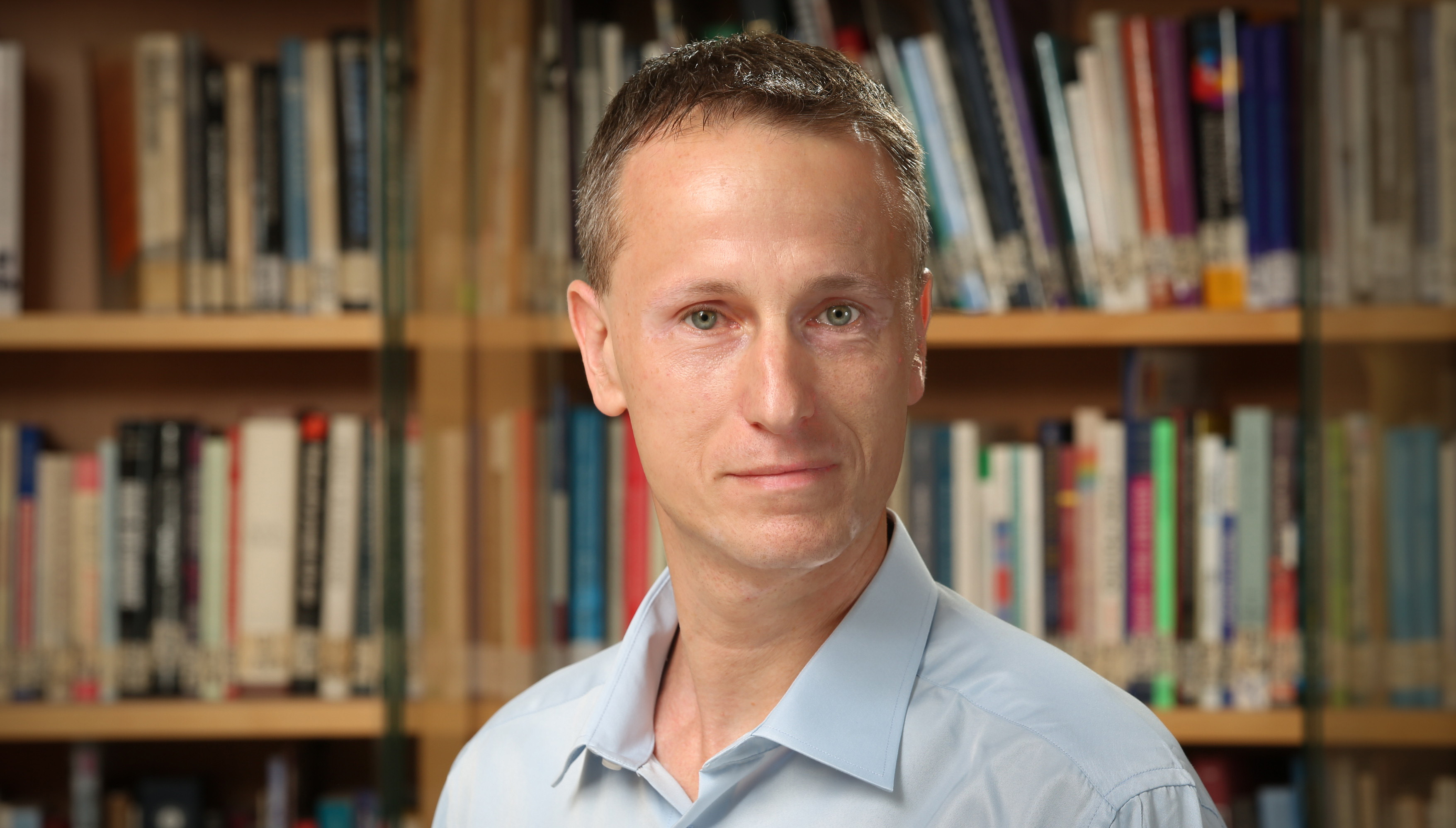
The study was conducted by Dr. Lydia Makrygianni, formaly a postdoctoral at Tel Aviv University and currently a researcher at Lancaster University in the UK. She led the research under the supervision of Prof. Iair Arcavi, a faculty member in the Astrophysics Department at TAU and Director of the Wise Observatory in Mitzpe Ramon.
Additional contributors included Prof. Ehud Nakar, Chair of TAU’s Astrophysics Department, and students Sara Faris and Yael Dgany from Prof. Arcavi’s research alongside multiple international collaborators. The results were published in the July 2025 issue of the Astrophysical Journal Letters.

Research team (Left to right): Sara Faris, Yael Dgany & Prof. Iair Arcavi
At the center of nearly every large galaxy lies a supermassive black hole, with mass millions to billions of times greater than that of the sun. One such black hole is located at the center of our own Milky Way Galaxy, and its discovery was recognized with the 2020 Nobel Prize in physics.
Still, much remains unknown about how these black holes form and influence their surroundings. Because they do not emit light, their presence is difficult to detect. In our galaxy, they are identified by the movement of nearby stars. But in distant galaxies, astronomers rely on rare, high-energy events to uncover their existence.
Once every 10,000 to 100,000 years, a star may wander too close to the black hole at the center of its galaxy and get ripped to shreds by its immense gravitating pull. Roughly Half of the star’s material is “swallowed” by the black hole, while the rest is ejected outward.
As the material falls in, it spirals in a circular motion, much like water going down a bathtub drain. Near the black hole, the rotating matter approaches the speed of light, heats up, and radiates intensely. For a few weeks to months, this flare “illuminates” the black hole, giving scientists a rare opportunity to observe its properties.
Yet strangely, many of these flares have not behaved as expected. Their brilliance and temperature have often been much lower than predicted, leaving researchers searching for explanations
According to the TAU-led team, the recently observed flare closely resembled AT 2022dbl, an earlier flare detected from the same location about two years prior.
This unusual repetition raises a new possibility: the first flare may have been caused by a partial disruption, in with the star was not fully destroyed and later returned for a second passage.
“The question now is whether we’ll see a third flare after two more years, in early 2026” says Prof. Arcavi. “If we see a third flare”, he continues, “it means that the second one was also the partial disruption of the star. So maybe all such flares, which we have been trying to understand for a decade now as full stellar disruptions, are not what we thought”.
If no third flare is observed, the second event may have been a full disruption. Whether or not a third flare occurs, the findings suggests that partial and full stellar disruptions may appear nearly identical, a prediction previously proposed by Prof. Tsvi Piran and his team at the Hebrew University of Jerusalem. “Either way”, adds Prof. Arcavi, “we’ll have to re-write our interpretation of these flares and what they can teach us about the monsters lying in the centers of galaxies”.

Research
Astounding Discovery at Tel Aviv University: Female moths decide where to lay their eggs based on sounds emitted by nearby plants
A scientific breakthrough at Tel Aviv University: A world-first study shows an acoustic interaction between plants and insects. In this study, the team focused on female moths and found that they make a critical decision—where to lay their eggs—based on sounds emitted by nearby plants. When plants emitted distress sounds, the female moths preferred healthy plants that were not emitting such sounds. These sounds are ultrasonic, beyond the hearing range of the human ear, but moths can hear them.
The study was conducted in the laboratories of Prof. Yossi Yovel from the School of Zoology and Prof. Lilach Hadany from the School of Plant Sciences and Food Security at TAU's Wise Faculty of Life Sciences. It was led by students Dr. Rya Seltzer and Guy Zer Eshel, in collaboration with scientists from the Plant Protection Institute at the Volcani Institute. The paper was published in the journal eLife.
This study follows the dramatic discovery published by the same researchers about two years ago, which generated worldwide interest: plants under stress emit sounds—at ultrasonic frequencies, above the range of human hearing, but detectable by many animals. The researchers state: “That discovery opened the door to extensive research on acoustic communication between plants and animals. In the present study, we began to explore this subject.”
Prof. Yovel explains: “After proving in the previous study that plants produce sounds, we hypothesized that animals capable of hearing these high-frequency sounds may respond to them and make decisions accordingly. Specifically, we know that many insects, which have diverse interactions with the plant world, can perceive plant sounds. We wanted to investigate whether such insects actually detect and respond to these sounds.”
Prof. Hadany adds: “We chose to focus on female moths, which typically lay their eggs on plants so that the larvae can feed on them once hatched. We assumed the females seek an optimal site to lay their eggs — a healthy plant that can properly nourish the larvae. Thus, when the plant signals that it is dehydrated and under stress would the moths heed the warning and avoid laying eggs on it? To explore this question, we conducted several experiments would the moths heed the warning and avoid laying eggs on it? To explore this question, we conducted several experiments.”
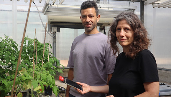
Prof. Yossi Yovel and Prof. Lilach Hadany
In the first experiment, aiming to isolate the auditory component from other plant features like color and scent, the researchers presented the female moths with two boxes: one contained a speaker playing recordings of tomato plants in a state of dehydration, while the other was silent. The moths showed a clear preference for the ‘noisy’ box, which they likely interpreted as a living plant (even if under stress). Conclusion: the moths do indeed perceive and respond to a playback of plant-emitted sounds. When the researchers neutralized the moths’ hearing organs, this preference disappeared and they chose both boxes equally — clear evidence that the preference was specifically based on listening to sounds, and not on other stimuli.
In the second experiment, the female moths were presented with two healthy tomato plants – one with a speaker playing sounds of a drying plant, and one that was silent. Again, they showed a clear preference – but this time for the silent plant, from which no distress sounds were heard, and therefore probably serves as a better site for laying eggs.

In another experiment, the moths again faced two boxes—one silent and the other containing male moths, which also emit ultrasonic sounds at a frequency similar to plant sounds. This time, the females showed no preference and laid their eggs equally on both boxes. The researchers concluded that when deciding where to lay their eggs, the females specifically respond to plant-emitted sounds — and not, for example, to sounds made by males.
The researchers conclude: “In this study, we revealed the first evidence for acoustic interaction between a plant and an insect. We are convinced, however, that this is just the beginning. Acoustic interaction between plants and animals doubtlessly has many more forms and a wide range of roles. This is a vast, unexplored field — an entire world waiting to be discovered.”

Research
Groundbreaking collaborative research leads to a novel mRNA-based vaccine targeting a lethal bacterial infection
Researchers from Tel Aviv University and the Israel Institute for Biological Research in Ness Ziona have used the platform developed for COVID-19 vaccines to create the world’s first mRNA-based vaccine against a deadly, antibiotic-resistant bacterium. In this groundbreaking study, the researchers tested the vaccine’s resistance to the virulent pathogen that causes the disease and were able to demonstrate 100% protection against infection in animal models. The researchers now hope that this technology can be used to combat other lethal bacteria as well.
The study was led by Tel Aviv University’s Vice President for Research and Development Prof. Dan Peer, a global pioneer in mRNA drug development and director of the Laboratory of Precision NanoMedicine at the Shmunis School of Biomedicine and Cancer Research. He worked alongside researchers from the Israel Institute for Biological Research — Dr. Uri Elia, Dr. Yinon Levy, Dr. Emmy Mamroud, and Dr. Ofer Cohen — as well as members of his own laboratory team: Dr. Edo Kon, Dr. Inbal Hazan-Halevy, and doctoral student Shani Benarroch. The study was featured on the cover of the prestigious journal Advanced Science.
The vaccine developed by the team from the Institute for Biological Research and Tel Aviv University is an mRNA-based vaccine delivered via lipid nanoparticles, similar to the COVID-19 vaccine. However, mRNA vaccines are typically effective against viruses like COVID-19 — not against bacteria like the plague.
Dr. Uri Elia explains: “Viruses rely on a host cell to survive and replicate. They infect the cell with an RNA molecule (mRNA) that contains instructions for making viral proteins. The virus uses the cell as a factory to replicate itself. In an mRNA vaccine, this molecule is synthesized and encased in a lipid nanoparticle that resembles human cell membranes. The nanoparticle fuses with the cell, the cell produces the viral proteins, and the immune system learns to recognize and defend against the actual virus upon exposure. Bacteria, however, are a different story: they produce their own proteins and do not rely on human cells. Moreover, due to the different evolutionary paths of humans and bacteria, their proteins are very different from ours.”
In 2023, the researchers developed a unique method for producing the bacterial protein within a human cell in a way that prompts the immune system to recognize it as a genuine bacterial protein and thus learn to defend against it. The researchers from Tel Aviv University and the Institute for Biological Research proved, for the first time, that it is possible to develop an effective mRNA vaccine against bacteria. They chose Yersinia pestis, the bacterium that causes bubonic plague — a disease responsible for deadly pandemics throughout human history. In animal models, the researchers demonstrated that it is possible to effectively vaccinate against the disease with a single dose.
Prof. Dan Peer: “In the previous study, we developed a vaccine for a form of plague transmitted through the skin — for example, via flea bites. In the current study, we chose a much more ambitious target: pneumonic plague, which spreads from person to person and causes respiratory illness — making it particularly difficult to develop a vaccine against. For this reason, we used two proteins — two antigens — to create the vaccine. We tested it on several animal model strains and found that, after two vaccine doses, we achieved 100% protection against pneumonic plague: the animals infected with the plague did not get sick at all. The success of the current study paves the way for a whole world of mRNA-based vaccines against other deadly bacteria.”
“The plague — a disease that killed about two-thirds of Europe’s population in the Middle Ages (‘The Black Death’) still resurfaces occasionally today, for example in Madagascar. So the potential for a pandemic still exists,” says Dr Uri Elia. “The disease is caused by a bacterium called Yersinia pestis, for which there is no approved vaccine in Western countries. This bacterium is highly contagious and extremely lethal, making it a serious threat. Moreover, this bacterium concerns us as a potential agent of bioterrorism. If one of our enemies tries to use it against us, we want to be prepared with a vaccine.”

Research
Two Orit books (the Torah of Beta Israel – Ethiopian Jewry) from the 15th century were discovered by the Orit Guardians program of TAU's Department of Biblical Studies.
A Rare Discovery: A traveling workshop of TAU's Orit Guardians program discovered two 15th-century Orit books – the oldest found to date in the possession of Beta Israel. The Orit Guardians Master's program was established about five years ago, with the primary goal of studying, preserving, and carrying on the Biblical heritage of Ethiopian Jewry. The workshop was held in collaboration with the Ethiopian Jewry Heritage Center and the National Library of Israel, which documented the books and established the Digital Archive of Beta Israel's Scriptures. Recently, these books were exposed at a special event held at ANU – Museum of the Jewish People, led by the Koret Center for Jewish Civilization (a collaboration between ANU and TAU), which supports and advances the Orit Guardians program.
The program's initiator, Prof. Dalit Rom-Shiloni from the Department of Biblical Studies, Chaim Rosenberg School of Jewish Studies and Archaeology, explains: "The Orit of Beta Israel includes the Five Books of the Torah, as well as the Books of Joshua, Judges, and Ruth. So far, we have documented four Orit books, including the two from the 15th century, as well as 13 other sacred books. All the sacred books of Ethiopian Jewry are written in Ge'ez, a language known only to the Kessim, and each manuscript has its own fascinating story. They have been passed down through generations from father to son, and some were given to Kessim by their teachers — Jewish monks who taught the sacred traditions in Ethiopia. The books were carefully guarded and preserved, with some of their owners even risking their lives to bring them to Israel. Today, most of these books are privately owned by Kessim and their families and used as “living books” in the prayer houses of Ethiopian Jewish communities across Israel. Until now, they were inaccessible to interested individuals of the general public, nor to the research world, and we intend to locate as many books as possible for preservation, digitization, and academic study."
To this end, a unique traveling workshop was held in June 2024, with participants including: Prof. Rom-Shiloni, anthropologist Prof. Erica Weiss, linguist Dr. Anbessa Teferra, and students from the Orit Guardians program — all from Tel Aviv University, alongside representatives of the Ethiopian Jewry Heritage Center and the National Library, as well as three international experts in ancient Ethiopian sacred texts: Prof. Loren Stuckenbruck (Ludwig Maximilian University of Münich), Dr. Sophia Dege-Müller and Ted Erho (University of Hamburg). The international experts examined and dated the books using palaeography — based on script forms. To their astonishment, they found that two of the Orit books were written as early as the 15th century — the oldest discovered so far in the hands of Beta Israel. Prof. Rom-Shiloni explains: "Our discovery is causing a stir among experts in the field worldwide. While we are familiar with similar Ethiopian texts from this period or even earlier, all of those are Christian texts, not Jewish. Now, for the first time, it has been revealed that Kessim from Beta Israel possess Orit books that are over 600 years old."
In total, the workshop yielded four Orit books — two from the 15th century and two from the 18th century, as well as 13 other sacred books from the 17th to 20th centuries. All the discovered books were documented with their owners’ consent and remain in their possession — so they can continue to serve as “living books” in their communities. The documentation now enables academic research and the establishment of a digital archive at the National Library.
Prof. Youval Rotman, Academic Director of the Koret Center and faculty member in the Department of Jewish History at Tel Aviv University, added: "This is an extraordinary finding. Discovering ancient manuscripts is rare, and when they are the oldest of their kind in existence, the find is all the more exceptional. This discovery was made thanks to the Orit Guardians' emphasis on studying the textual knowledge and interpretive tradition preserved and orally transmitted over centuries within the various Beta Israel communities. The uniqueness of the program lies not only in mapping manuscripts and training students for their research but also in doing so as an integral part of the knowledge preserved within the community — thereby continuing and expanding it. The young researchers form personal connections and earn the trust of the Kessim as successors to the tradition and oral interpretation. In doing so, they connect communal-social knowledge to academic knowledge — and this is our great pride. The program unearths hidden treasures that have so far dwelt within the four walls of local synagogues, then documents and studies them and makes them accessible. Imagine a situation in which the great Bible commentaries were passed down orally through generations."
Prof. Rom-Shiloni concludes: "Through the traveling workshop of the Orit Guardians program we discovered 17 sacred books of Beta Israel held by Kessim across Israel and still used as “living books” in the prayer houses of Ethiopian Jewry. Among them, we discovered two Orit books — the Torah of Beta Israel — written in the 15th century, the earliest found so far in the hands of Jewish Kessim.
Alongside the excitement, we believe our discovery is only the tip of the iceberg. It is likely that many more sacred books of Beta Israel are held by families and Kessim around the country, and we will continue searching for them. It is important to emphasize that all manuscripts located (like those found through the workshop) will remain in the possession of their owners, while being photographed and documented to make them accessible to interested members of the community, the broader public, and researchers in Israel and around the world. Another, especially urgent task of the Orit Guardians is documenting the scholarly oral traditions of the Kessim in Ethiopia, which includes translation from Ge’ez to Amharic and interpretation of the Orit and other holy books. This heritage, transmitted only orally from generation to generation, has never been set down in writing. Today, only 18 senior Kessim, who were trained in Ethiopia and hold this knowledge, remain active in Israel, and they are aging. If we do not act quickly, we might lose this precious cultural treasure."

Research
New study by the Moshe Dayan Center at Tel Aviv University examines the views of Arab citizens in Israel – after the war with Iran
A new study by the Konrad Adenauer Program for Jewish-Arab Cooperation at TAU's Moshe Dayan Center finds that a large majority (73.2%) of Israel's Arab citizens support the inclusion of an Arab party in the government that will be formed after the next elections. In addition, the turnout of Arab voters is expected to increase.
A large majority (73.2%) support the participation of an Arab party in the next government: 41.8% support joining any government formed, and 31.4% support joining a center-left government. If elections for the Knesset were held today, the expected voter turnout in Arab society would be 57%, slightly higher than the 53.2% turnout in the 25th Knesset elections held in November 2022.
The war between Israel and Iran did not change the priorities of Arab citizens regarding fundamental political issues such as joining the government or support for Arab-Jewish political partnership. However, some impact is evident in the definition of personal identity.
Most of the Arab public (66% of respondents) believe in political cooperation between Arabs and Jews in Israel, but only 40.2% believe that the Jewish public actually supports such cooperation.
A large majority of the Arab public (75.4%) report a low sense of personal security. The two main factors negatively affecting their mood are the high incidence of violence in Arab communities (41.9%) and the ongoing war in Gaza (37.6%). At the same time, 64% of survey participants report that their financial situation is relatively good.
54% of survey respondents stated that the most important issue for the Arab public today is addressing the problem of violence and crime. A significant portion (23.2%) said that ending the war in Gaza is the most important issue.
The personal identity of Israel's Arab citizens includes three main components: Arab identity (36.2%), Israeli citizenship (30.3%), and religious affiliation (21.4%). For a relatively small portion of this public (9.7%), their Palestinian identity is the most important.
The study was initiated by the Konrad Adenauer Program for Jewish-Arab Cooperation, operating under the German Konrad Adenauer Stiftung at the Moshe Dayan Center for Middle Eastern and African Studies at Tel Aviv University.
Dr. Arik Rudnitzky, Director of the Konrad Adenauer Program for Jewish-Arab Cooperation at the Moshe Dayan Center: “A large, solid majority of Israel's Arab citizens support political partnership between Arabs and Jews, as well as the inclusion of an Arab party in the government that will be formed after the next elections. The importance of this political standpoint cannot be overstated".
"It should be understood against the backdrop of a harsh reality in which the mood in the Arab public is low due to the rampant violence in Arab communities, the negative effects of the war in Gaza, and also the recent brief war with Iran, which starkly exposed the severe lack of protective facilities in Arab communities. Nevertheless, and contrary to expectations, Israel's Arab citizens refuse to despair and look soberly at the day after the war. According to this survey, voter turnout of Arab citizens in the next Knesset elections will be slightly higher than in the elections of November 2022. Opinions are divided on whether the precedent of the inclusion of Arab party Ra’am in the Bennett-Lapid government (2021-22) was successful, but even those who criticize MK Mansour Abbas (Head of Ra'am) for this move do not necessarily oppose the attempt itself — rather, they believe Ra’am could have achieved more. Thus, precisely in the context of the longest and harshest war in the history of the Israeli-Palestinian conflict, the survey points to the hope residing in the hearts of Arab citizens for establishing a political partnership between Arabs and Jews in the day after the war".
Dr Arik Rudnitzky


