


Political orientation can be predicted by measuring brain activation while watching campaign-ads


Research
Political orientation can be predicted by measuring brain activation while watching campaign-ads
A first-of-its-kind study scanned the brains of dozens of politically involved participants while they watched campaign-ads and speeches by parties from both ends of the political spectrum, just before one of the last rounds of elections. The participants, half right-wing and half left-wing, were scanned using magnetic resonance imaging (fMRI), a method that measures brain activation. Surprisingly, political-dependent differences in the brain response emerged already in early brain regions, such as regions involved in vision and hearing, and in fact the response in these regions was enough to predict an individual's political views.
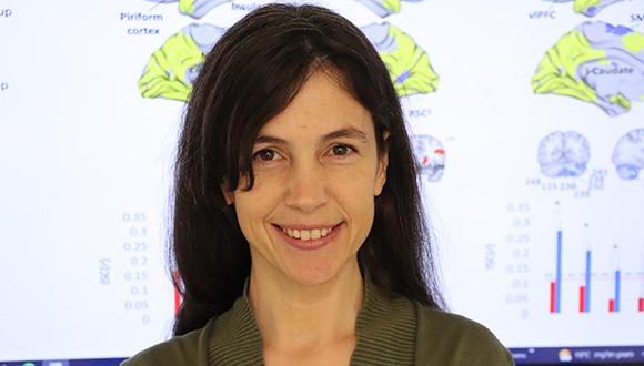
The study was led by Noa Katabi, a research student in the lab of Dr. Yaara Yeshurun in The School of Psychological Sciences and the Sagol School of Neuroscience. The study was published in the Journal of Neuroscience.
During the study, participants watched video-clips, including a neutral (in terms of political characteristics) video-clip and different political campaign-ads and political speeches by politicians from both blocs, Right and Left. The researchers were surprised to discover widespread partisanship-dependent brain activation and synchronization when Right-wing individuals watched the videos of their political bloc, or when Left-wing individuals watched the videos of left-wing politician.
Interestingly, the researchers found that such partisanship-dependent differences in brain synchronization was not limited to "higher" areas of the brain, associated with interpretation and abstract thinking, as was previously found. Rather, these differences occurred already in regions responsible for sight, hearing and even touch.
"This is the first study to show political-dependent brain activity in early sensory and motor areas, and it can be said that at the most basic brain level, rightists and leftists in Israel literally (and not just metaphorically) don't see and hear the same things." Dr. Yaara Yeshurun
Dr. Yaara Yeshurun
"The research clearly showed that the more the subjects were politically aligned with a certain group, the more their brain response was synchronized, including in motor and somatosensory areas, that is, those areas of the brain that are active when we move or feel things with our senses," explains Dr. Yeshurun. "In fact, just by the brain’s response in these primary sensory areas we could tell if a certain individual was left or wight wing. Intriguingly, it was not necessary to examine the activity in 'higher' brain areas - areas that are involved in understanding why a certain character did something, or what that character thinks and feels - to predict participants' political views, it could even be done by examining an area of the brain that is responsible for seeing or hearing.”
The researchers think that this surprising finding is due to the fact that the participants they chose were politically involved, and also due to the timing of the experiment - a few weeks before the elections, when the political atmosphere in Israel was very present and emotional.
"This is the first study to show political-dependent brain activity in early sensory and motor areas, and it can be said that at the most basic brain level, rightists and leftists in Israel literally (and not just metaphorically) don't see and hear the same things. I think that if we try to understand how people who hold opposite political views to ours experience the world, we might be able to conduct a slightly more effective public discussion that can hopefully attenuate the current political polarization,” adds Dr. Yeshurun.

Right or left? "If we try to understand how people who hold opposite political views to ours experience the world, we might be able to conduct a slightly more effective public discussion (…)"

Research
Findings contradict prevalent belief that people on the autism spectrum are 'indifferent to pain'
A new study examined the pain perception among people with autism and found that they experience pain at a higher intensity than the general population and are less adaptable to the sensation. This finding is contrary to the prevalent belief that people with autism are supposedly 'indifferent to pain'. The researchers expressed the hope that the findings of their study will lead to more appropriate treatment on the part of medical staff, caregivers, and parents toward people with autism, who do not always express the experience of pain in the usual way.
The study was funded by the Israel Science Foundation and was led by four researchers: Dr. Tami Bar-Shalita of the Sackler Faculty of Medicine at Tel Aviv University who initiated the study, Dr. Yelena Granovsky of the Technion and Rambam Medical Center, and Prof. Irit Weissman-Fogel and Prof. Eynat Gal of the University of Haifa. This study constitutes a framework for the theses of PhD students Tzeela Hofmann and Mary Klingel-Levy, and three articles based on it have already been published or approved for publishing. The present study has been published in the prestigious PAIN journal.
"We know that self-harm could stem from attempts to suppress pain, and it could be that [people with autism] hurt themselves to activate, unconsciously, a physical mechanism of 'pain inhibits pain'." Dr. Tami Bar-Shalita
"Approximately 10% of the general population suffer from sensory modulation dysfunction, which means sensory hypersensitivity at a level that compromises normal daily functioning and quality of life. These people have difficulty, for example, ignoring or adapting to buzzing or flickering of fluorescent lights, humming of air conditioners or fans, or the crunching of popcorn by someone sitting next to them in the cinema," explains Dr. Bar-Shalita.
"In previous studies in the lab we found that these people suffer from pain more than those without sensory modulation dysfunction. Since it is known that sensory modulation dysfunction occurs in people with autism at a rate of 70-90%, it constitutes a criterion for diagnosing autism, and is associated with its severity. We were interested in exploring pain perception in autism, so we asked: do people with autism hurt more than the general population? This question was hardly studied in the lab before we got started."
According to the researchers, for many years the prevalent opinion was that 'people with autism hurt less' or that they were 'indifferent to pain'. Actually, 'indifference to pain' is one of the characteristics presented in the current diagnostic criteria of autism.
The proof of this was, supposedly, their tendency to inflict pain on themselves by self-harm.
Dr. Bar-Shalita: "this assumption is not necessarily true. We know that self-harm could stem from attempts to suppress pain, and it could be that they hurt themselves to activate, unconsciously, a physical mechanism of 'pain inhibits pain'."
"The results of our study indicate that in most cases, the sensitivity to pain of people with autism is higher than that of most of the population, while at the same time they are unsuccessful at effectively suppressing painful stimuli." Dr. Tami Bar-Shalita

Dr. Tami Bar-Shalita
This study is a laboratory pain study approved by the ethics committee of the academic institutions and Rambam Medical Center. The study included 52 adults with high-functioning autism (HFA) and normal intelligence – hitherto the largest reported sample in the world in studies on pain among people with autism. The study made use of psychophysical tests to evaluate pain, commonly used in the area of pain study. These methods examine the link between stimulus and response, while the researcher, using a computer, controls the duration and intensity of stimulus and the examinee is asked to rank the intensity of the pain felt by him on a scale of 0 to 100.
The findings have proven beyond doubt that people with autism hurt more. Furthermore, their pain suppression mechanism is less effective.
The researchers conducted a variety of measurements, aimed among other things at examining whether the hypersensitivity to pain derives from a sensitized nervous system or from suppression of mechanisms that are supposed to enable adjustment and, over time, reduce the response to the stimulus. They found that in the case of people with autism, it is a combination of the two: an increase of the pain signal along with a less effective pain inhibition mechanism.
Dr. Bar-Shalita concludes: "our study constituted a comprehensive, in-depth study of the intensity of pain experienced by people with autism. The prevalent belief was that they are supposedly 'indifferent to pain', and there are reports that medical and other professional staff treated them accordingly. The results of our study indicate that in most cases, the sensitivity to pain of people with autism is higher than that of most of the population, while at the same time they are unsuccessful at effectively suppressing painful stimuli. We hope that our findings will benefit the professionals and practitioners handling this population and contribute to the advancement of personalized treatment."
In additional articles soon to be published, the researchers have examined the brain activity of people with autism during pain stimuli, and sub-groups within this population concerning their perception of pain.

Research
Findings may contribute to development of treatments to enable normal expression of genes essential for brain development in people suffering from the syndrome
Williams syndrome is a relatively rare, multisystem genetic syndrome that causes disorders in brain development. A new study by the Tel Aviv University and Hebrew University found that abnormal processes lead to disruption in the expression of genes essential for brain development in people suffering from the syndrome. The researchers believe their findings may contribute to the future development of targeted treatments that will enable normal expression of the affected genes identified in the research.
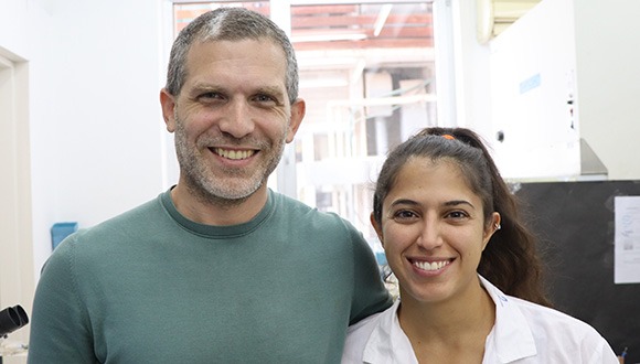
The research was led by Dr. Boaz Barak from the School of Psychological Sciences and the Sagol School of Neuroscience at Tel Aviv University and Dr. Asaf Marco from the Faculty of Agriculture, Food and Environment of the Hebrew University. Also participating in the research were Dr. Sari Trangle, Mr. Gilad Levy and Ms. Ela Bar from Dr. Barak's laboratory, and Dr. Tali Rosenberg and Ms. Hadar Parnas from Dr. Marco's laboratory. The research findings were published in the prestigious journal Molecular Psychiatry from the Nature publishing group.
"We wanted to examine whether the Williams Syndrome is also characterized by defects in the genomes contained in brain cells which prevent the proper expression of essential genes." Dr. Boaz Barak
Dr. Barak: “Williams syndrome is a rare, multisystem genetic syndrome that includes disorders in brain development that lead to heightened social interactions, mental retardation, and other characteristic features. Past research has revealed that twenty-five genes are missing from the DNA on chromosome number seven of people with Williams syndrome, and the study of the syndrome to date has mainly focused on those missing genes and their functions."
"We wanted to examine whether the syndrome is also characterized by defects in the genomes contained in brain cells which prevent the proper expression of essential genes. Specifically, we asked: ‘Is it possible that certain genes are not expressed properly in the brains of people with Williams syndrome due to the phenomenon of methylation - when a molecule known as a 'methyl group' is located on a certain gene that is present in the genome, preventing it from expressing itself properly?".
To illustrate the phenomenon of the missing genes, Dr. Barak took an instruction book in which some of the pages were torn out. As a result of the missing pages, anyone following the instructions would make mistakes. Similarly, hiding some of the letters in the pages left in the book with a black marker would result in instructions being corrupted, just like methylation on an existing gene disrupts its expression.
Methylation is in many cases a normal mechanism in the cells of the body, as its role is to prevent expression of certain genes when appropriate. However, when there are disruptions in the correct application of methylation, the abnormal expression of the genes may lead to impairments in cell function, and subsequently cause damage to various organs, including to normal brain development.
Dr. Boaz Barak from the School of Psychological Sciences and the Sagol School of Neuroscience at Tel Aviv University
The researchers examined human brain tissues taken from adults with and without Williams syndrome who died of causes unrelated to the syndrome and donated their brains to science.
“We focused on samples from the frontal lobe, the area of the brain that is responsible for brain functions such as cognition and decision-making," Dr. Barak explained. "In a previous study, we located in this area damage to the characteristics of the nerve cells and the cells that support nerve cell activity in people with Williams syndrome. In this study, we examined all the genes in all the cells of the frontal lobe to determine whether there are genes in people with Williams syndrome that have undergone abnormal methylation processes, i.e., increased or decreased gene silencing compared to a brain with typical development.”
"We uncovered significant information about the defective expression of genes in people with Williams syndrome. While these genes are fully present in the genome of the brain cells, until now it was not known that these abnormally regulated genes are involved in the syndrome." Dr. Asaf Marco.
The researchers found that indeed in people with Williams syndrome abnormal methylation does exist in this area of the brain, resulting in disruption of the normal expression of many genes related to the normal development of the brain's neural functions, such as regulation of social behavior (people with Williams syndrome are known to be overly friendly), cognition, plasticity of the brain, and cell survival.
“We uncovered significant information about the defective expression of genes in people with Williams syndrome. While these genes are fully present in the genome of the brain cells, until now it was not known that these abnormally regulated genes are involved in the syndrome," says Dr. Marco.
"Building on our findings, it will be possible to focus future efforts on the development of targeted treatments that will reach the disrupted sites that we identified in the study in order to 'correct' the defective expressions." Dr. Boaz Barak
"In addition, one of our main findings is that the disruptions in methylation do not have to appear near the gene whose function is impaired, and sometimes the disruptions are located far away from it. This information is critical because it allows us to better understand the spatial organization of DNA and its effect on gene control."
He adds that, "since we know of enzymes that are able to remove or add methyl molecules, the next challenge will be to precisely direct those enzymes to the disrupted sites identified in our research, with the aim of allowing the genes to be properly expressed.”
Dr. Barak concludes: "Our research revealed new factors related to the disabilities that characterize Williams syndrome. Instead of focusing on the effects of the missing gene, as has been done until now, we shed light on many more genes that are expressed in a defective manner. Building on our findings, it will be possible to focus future efforts on the development of targeted treatments that will reach the disrupted sites that we identified in the study in order to 'correct' the defective expressions.”

Research
In a scientific first, a robot can “smell” using a biological sensor
After having developed a robot that hears through the ear of a locust, researchers from Tel Aviv University have succeeded in equipping a robot with the sense of smell, using a biological sensor. The sensor sends electrical signals as a response to the presence of a nearby odor, which the robot can detect and interpret. The researchers successfully connected the biological sensor to an electronic system and using a machine learning algorithm, were able to identify odors with a level of sensitivity 10,000 times higher than that of a commonly used electronic device. The researchers say "The sky's the limit," and believe that this technology may also be used in the future to identify explosives, drugs, diseases, and more.
“Man-made technologies still can’t compete with millions of years of evolution. One area in which we particularly lag behind the animal world is that of smell perception (…) When they want to check if a passenger is smuggling drugs [at the airport], they bring in a dog to sniff him." Dr. Ben Maoz and Prof. Amir Ayali
The biological and technological breakthrough was led by doctoral student Neta Shvil of Tel Aviv University’s Sagol School of Neuroscience, Dr. Ben Maoz of the Fleischman Faculty of Engineering and the Sagol School of Neuroscience, and Prof. Yossi Yovel and Prof. Amir Ayali of the School of Zoology and the Sagol School of Neuroscience. The results of the study were published in the prestigious journal Biosensor and Bioelectronics.
Dr. Maoz and Prof. Ayali explain: “Man-made technologies still can’t compete with millions of years of evolution. One area in which we particularly lag behind the animal world is that of smell perception (…) When they want to check if a passenger is smuggling drugs [at the airport], they bring in a dog to sniff him."
"In the animal world, insects excel at receiving and processing sensory signals. A mosquito, for example, can detect a 0.01 percent difference in the level of carbon dioxide in the air. Today, we are far from producing sensors whose capabilities come close to those of insects.”
The researchers point out that, in general, our sensory organs, such as the eye, ear and nose – as well as those of all other animals – use receptors that identify and distinguish between different signals. Then, the sensory organ translates these findings into electrical signals, which the brain decodes as information. The challenge of biosensors is in the connection of a sensory organ, like the nose, to an electronic system that knows how to decode the electrical signals received from the receptors.
Dr. Ben Maoz and doctoral student Neta Shvil
“Nature is much more advanced than we are, so we should take advantage of that." Dr. Ben Maoz.
“We connected the biological sensor [to the electronic system] and let it smell different odors while we measured the electrical activity that each odor induced," explains Prof. Yovel, Kadar Family Award for Outstanding Research recipient. "The system allowed us to detect each odor at the level of the insect’s primary sensory organ."
"Then, in the second step, we used machine learning to create a ‘library’ of smells. In the study, we were able to characterize 8 odors, such as geranium, lemon and marzipan, in a way that allowed us to know when the smell of lemon or marzipan was presented. In fact, after the experiment was over, we continued to identify additional different and unusual smells, such as various types of Scotch whiskey. A comparison with standard measuring devices showed that the sensitivity of the insect’s nose in our system is about 10,000 times higher than the devices that are in use today.”
“Nature is much more advanced than we are, so we should take advantage of that," says Dr. Maoz. "The principle we have demonstrated can be used and applied to other senses, such as sight and touch. For example, some animals have amazing abilities to detect explosives or drugs; the creation of a robot with a biological nose could help us preserve human life and identify criminals in a way that is not possible today. Some animals can detect diseases. Others sense earthquakes. The sky is the limit.”
What's next? The researchers plan to give the robot a navigation ability to allow it to localize the odor source and later, its identity.

Will he be able to retire soon? A working dog searches for hazardous materials at the airport

Research
Israeli researchers find that medical clowns contribute significantly to the achievement of medical therapeutic goals
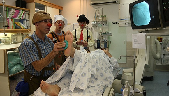
You see them stroll around in the hospitals' toughest wards with their red noses, colorful clothes, and unwavering smiles, spreading laughter and cheerfulness wherever they go. They are the medical clowns: trained professionals whose goal is to change the hospital environment through humor.
A new study tested and categorized the skills of medical clowns and found that their importance goes far beyond contributing to a patient's good mood. The researchers identified 40 different skills of medical clowns, including establishing an emotional connection and creating a personal relationship with the patient, expressing the patient's frustrations and difficulties to the medical staff, increasing the patient’s motivation to adhere to medical treatment, distracting the patient from pain, and creating a joyful atmosphere.
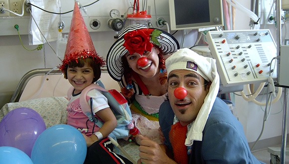
Medical clowns working alongside other therapists (Photo: The Dream Doctors Project, Medical Clowning in Action)
The research was conducted under the leadership of Prof. Orit Karnieli-Miller, with Dr. Lior Rosenthal, both from the Department of Medical Education at TAU's Sackler Faculty of Medicine, in collaboration with Ms. Orna Divon-Ophir, Dr. Doron Sagi, Prof. Amitai Ziv and Ms. Liat Pessach-Gelblum from the Israel Center for Medical Simulation (MSR). The study was published in Qualitative Health Research, a leading journal in the field of health.
The researchers show that not only do medical clowns help the patients and their family members, but also the medical team and the achievement of treatment goals.
Through use of different communication skills, clowns make it easier for the patient to cooperate with various treatments. The medical clowns work in a team with other therapists, know how to intervene and help whenever an argument or crisis should arise to advance treatment.
"From the moment they enter the room, the clowns form a bond with the patients, strengthen them, and give them power and status within the medical system." Prof. Orit Karnieli-Miller.
Studies conducted throughout the years have shown the clowns’ positive influence on the hospital environment through humor, as well as helping patients deal with pain. However, no studies have empirically mapped the skills they use and their therapeutic goals to help understand their “secret magic.” In addition, there was a lack of broad understanding of how clowns can help children, teenagers, and their parents in various challenging situations of distress and difficulty, as well as how they can help patients and medical teams achieve treatment goals. This lack of appreciation of the potential benefits of utilizing the services of medical clowns meant that patients and medical teams would occasionally be reluctant to cooperate with them.
As part of the new study, the researchers focused on qualitative, in-depth systematic identification of the skills of medical clowns through observation and analysis of their actions in challenging encounters with adolescents, parents, and medical staff.
Medical clowns help patients and medical teams achieve treatment goals (Photo: The Dream Doctors Project, Medical Clowning in Action)
The team analyzed videotaped sessions of medical clowns in various simulated situations and conducted in-depth interviews with expert medical clowns. The researchers identified 40 different skills used by the medical clowns to achieve four therapeutic goals:
1) building a relationship and connecting to the needs and desires of the patients
2) dealing with emotions and difficulties
3) increasing the patient’s motivation to adhere to the treatment plan
4) increasing the patient’s sense of control and providing encouragement to patients
The clowns examined in the study were trained and recruited by the "Dream Doctors Project”, a non-profit association that employs medical clowns as part of the paramedical system in Israeli hospitals, and trains them to work within multi-disciplinary teams. The Tel Aviv University researchers collaborated with the Israel Center for Medical Simulation (MSR), which created a simulation-based workshop focused on developing the skills of experienced medical clowns.
"From the moment they enter the room, the clowns form a bond with the patients, strengthen them, and give them power and status within the medical system," explains Prof. Karnieli-Miller. "They do this through an initial connection to the patients’ voice, and even to the patients’ reluctance to implement therapeutic recommendations - an emotional connection that often results in the patient changing their position and cooperating with the medical staff."
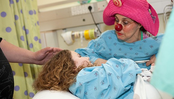
Providing the patient with an increased sense of control and courage to face their challenges (Photo: The Dream Doctors Project, Medical Clowning in Action)
According to Prof. Karnieli-Miller the medical system is hierarchical, and it is not always easy for patients to navigate. Therefore, one of the skills of medical clowns is to place themselves in the lowest position in the medical setting. By doing so, they empower the patients by giving them a sense of power and control, including the choice of whether to allow the clown to enter the room as well as to dictate the nature of the patient’s role vis-à-vis that of the clown. This provides the patient with an increased sense of control and courage to face their challenges.
The researchers emphasize that the clowns are very aware of the emotional difficulty associated with staying in a hospital and dealing with an illness. To help deal with these issues, the clowns sometimes distract the patient by using props, humor, and imagination. Other skills include allowing the patient to direct their frustrations towards them, away from medical staff or parents.
Depending on the situation the clowns may also use a comforting touch, soothing music, empathetic listening, or a reinforcing statement to provide an environment where the patient feels comfortable to express their feelings. A patient’s ability to gain legitimacy is important and is strengthened by the clowns.
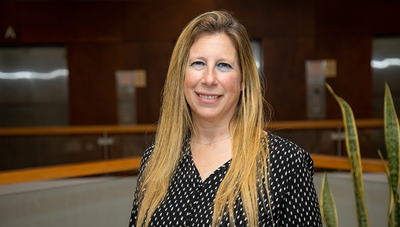
Prof. Orit Karnieli-Miller
"Mapping the skills and goals of the medical clowns improves their understanding of their role and may help other health professionals appreciate their work methods and the benefits of incorporating these methods into their own practices when faced with similar challenges" Prof. Orit Karnieli-Miller.
"Mapping the skills and goals of the medical clowns improves their understanding of their role and may help other health professionals appreciate their work methods and the benefits of incorporating these methods into their own practices when faced with similar challenges,” adds Prof. Karnieli-Miller.
"This research is important because it allows the clowns to enhance their training program and refine their diverse skills to achieve the various therapeutic goals appropriate for different patients, as well as helping health professionals collaborate with the medical clowns. If professionals in the healthcare field gain a clear understanding of how and when to cooperate with the medical clowns, they will be able to help patients overcome challenges, and at the same time they may be more tolerant of the clowns' ‘disruption’ of the hospital care regimen. This appreciation of the clowns’ contribution will provide the clowns with the time and space to connect with patients and help and encourage patients to become more active participants in their treatment plan,” she concludes.


