


CPAT, a groundbreaking Tel Aviv University development, offers promising results with sustained improvement months after treatment


Research
CPAT, a groundbreaking Tel Aviv University development, offers promising results with sustained improvement months after treatment
Attention Deficit Hyperactivity disorder (ADHD) is one of the most common mental disorders affecting children. Symptoms of ADHD include inattention, hyperactivity, and impulsivity, and the disorder is considered a chronic and debilitating disorder that affects many aspects of an individual's life, including academic and professional achievements, interpersonal relationships, and daily functioning.
Tel Aviv University has developed a new treatment called Computerized Progressive Attention Training (CPAT), which has shown remarkable efficacy in alleviating symptoms of Attention Deficit Hyperactivity Disorder (ADHD) among students. In fact, a notable 33% of students experienced significant improvements in their symptoms when undergoing CPAT, surpassing the improvement in symptoms of only 11% of the students who participated in a parallel protocol involving mindfulness training. During this mindfulness training, students practiced a specific form of meditation designed to mitigate their attention difficulties. Importantly, the benefits of CPAT also outshone those of drug treatments like Ritalin, as the improvements persisted for up to four months after the completion of the treatment protocol.
The study was the doctoral dissertation of Dr. Pnina Stern, under the guidance of Prof. Lilach Shalev-Mevorach of The Jaime and Joan Constantiner School of Education at Tel Aviv University. The encouraging results of the study were recently accepted for publication in the Journal of Attention Disorders.
“We developed the CPAT system years ago, and it produced good results in previous studies that we conducted, mainly in children," explains Prof. Shalev-Mevorach. "Furthermore, in the only study that we conducted in adults with ADHD, positive findings were obtained, but without indications of ‘far transfer,’ meaning an improvement in functions for which participants were not directly trained in the treatment.”
According to Prof. Shalev-Mevorach, it is challenging for researchers to make scientific claims about the effectiveness of non-medication treatments because it is difficult to compare them to a "non-medication placebo." In other words, when studying non-medication treatments, it's hard to distinguish the effects of the treatment itself from other factors like the attention participants receive during training sessions or the effort they put into the research. This makes it complex to determine the true impact of non-medication interventions.

Prof. Lilach Shalev-Mevorach
In the current study, the team of researchers tried to resolve this by employing a research design that included two control groups: a regular control group, which performed the various assessment tasks at two points in time without any intervention as part of the research (the passive control group) and a second control group that participated in mindfulness training sessions under the guidance of a professional instructor. This type of training has yielded positive results in previous studies in people with ADHD.
For the experiment 54 students, male and female, diagnosed with ADHD were recruited from Tel Aviv University and other academic institutions. The subjects were blindly divided into three groups: a zero-intervention control group, a mindfulness group and a CPAT group.
Participants in the CPAT and the Mindfulness groups attended two-hour long group meetings on the University campus once a week, where the CPAT group received Computerized Progressive Attention Training and the mindfulness group received training from a certified mindfulness instructor.
Before and after the intervention protocol, the participants of the three groups performed a comprehensive series of assessment tests: standard computerized tests to assess attention functions, behavioral assessment questionnaires (self-reported ADHD symptoms), and mindfulness questionnaires (self-reported feelings such as stress, anxiety and well-being). In addition, a novel measurement was used for this intervention study, whose participants were, as mentioned, higher-education students: they were asked to read a text from a scientific article while their eye movements were tracked by an eye-tracker. The indices produced using the eye-tracking system made it possible to identify a pattern of inattentive reading, which was used as a measure of reading efficiency in an academic context. Finally, the participants filled out a questionnaire regarding their academic difficulties.
Prof. Shalev-Mevorach says the results were very positive: “We saw improvements in the attention functions themselves, that is, ‘near transfer,’ for example in sustained attention, the ability to remain attentive for a long period of time, and in attention control, the ability to delay a routine response. But the main thing, is that we saw significant improvements in the participants’ daily and academic functioning, such as reduced repeated reading while reading a scientific article. Furthermore, the improvements in these attention functions were connected to the reduction in behavioral symptoms of ADHD and in repetitive reading."
"In other words, the CPAT trained the attention mechanisms themselves, and their improvement was related to the improvement achieved in behavioral symptoms and reading patterns. 33% of the participants who received the CPAT protocol showed a significant improvement in ADHD symptoms, compared to only 11% of those who underwent the mindfulness protocol. The improvements obtained were preserved in the testing that was carried out about four months after the end of the intervention protocol.”
Prof. Shalev-Mevorach notes that the effects of stimulant drugs (psychostimulants) such as Ritalin and Concerta are ‘on-off’: patients who take Ritalin daily enjoy significant improvements, but when they stop the treatment, the improvements fade, and they return to the starting point. She says the researchers wish to bring about "a profound change in basic attention functions, a change that will be significant in the long term, as an additional option alongside medication, and of course as an alternative to drug treatment in cases in which it isn’t applicable.”

Research
The role of hope in supporting mental health
The role of hope in supporting mental health is not sufficiently understood among relevant professionals, according to Dr. Dorit Redlich Amirav of TAU’s Department of Occupational Therapy, Steyer School of Health Professions, Sackler Faculty of Medicine.
“Hope is similar to the air we breathe,” says Redlich Amirav. “Air is taken for granted in our daily life until we are suffocating and struggling to breathe.”
Redlich Amirav studies how different groups implement hopeful thinking and improve mental well-being through meaningful occupations. Through her findings, she aspires to help mental health professionals to integrate concepts of hope into their research and treatment and, in the long run, provide a longer-lasting and greater impact on each patient’s holistic well-being.
"Hope is similar to the air we breathe. Air is taken for granted in our daily life until we are suffocating and struggling to breathe." - Dr. Dorit Redlich Amirav
In new research published in Qualitative Health Research, she investigated the cross-generational transmission of hope. Redlich Amirav cites one of her female participants who was forced by her grandfather to quit school in sixth grade. She felt her hope diminish but stated that her hopeless personal circumstances led her to put more of an emphasis on the importance of education and studying with her own two daughters who both graduated from university.
Other participants displayed this particular kind of cross-generational hope. For example, a mother told Redlich Amirav about her father, who was a violinist until the Nazis broke his fingers. The mother internalized this trauma in a negative way, but all four of her own children play instruments and one of them is an opera singer. She inadvertently conveyed how hope and music are intertwined for them and their heritage.
In 2019, Redlich Amirav was appointed director of the Israeli chapter of the International Hope Barometer. She says that it came just in time: hope became a key factor in successfully adapting to the trials and tribulations of the pandemic. During the lockdowns, she says that people found meaning in new ways of interacting; specific trends point to the importance of goal-directed behavior in increasing hope.
Source: TAU Review

Research
New study reveals that brain's coordination between hippocampus and cortex during sleep boosts memory consolidation, offering hope for people with memory impairments
While a good night's sleep is known to be critical for the consolidation of long-lasting memories, so far there has been little evidence regarding the precise processes at work during human sleep. A breakthrough study demonstrated for the first time that long-lasting memories are consolidated in the human brain through communication between the hippocampus and the cerebral cortex during sleep. Moreover, the researchers found that by inducing deep-brain stimulation during sleep they can improve memory consolidation. They believe intervention during sleep represents a unique approach that can be further developed in the future to provide hope for people with memory impairments such as dementia.
The unique study, which was published in the leading journal Nature Neuroscience, involved an international collaboration led by Dr. Maya Geva-Sagiv (today at UC Davis). The study was a collaboration between the laboratories of Prof. Yuval Nir from the Sackler Faculty of Medicine, Department of Biomedical Engineering at The Iby and Aladar Fleischman Faculty of Engineering, and Sagol School of Neuroscience at Tel Aviv University, and Prof. Itzhak Fried from the Department of Neurosurgery at UCLA and the Sackler Faculty of Medicine at Tel Aviv University.
"Intervention during sleep represents a unique approach that can be further developed in the future to provide hope for people with memory impairments such as dementia." - Prof. Yuval Nir

The researchers (from left to right): Dr. Maya Geva-Sagiv, Prof. Yuval Nir and Prof. Itzhak Fried
"This study was made possible by a rare group of 18 patients with epilepsy at the UCLA Medical Center," says Prof. Nir. "Prof. Fried implanted electrodes in these patients' brains to try and pinpoint the areas that cause their epileptic seizures, and they volunteered to take part in a study investigating the effects of deep-brain stimulation during sleep. Close work with expert neurologists led by Prof. Dawn Eliashiv at UCLA enabled our team to integrate advanced brain stimulation in the research. Thus, we were able to test, for the first time in humans, the long-held hypothesis - that coordinated activity of the hippocampus and cerebral cortex during sleep is a critical mechanism in consolidating memories."
"Moreover, we improved memory consolidation through a special stimulation protocol that enhanced synchronization between these two areas in the brain. Intervention during sleep represents a unique approach that can be further developed in the future to provide hope for people with memory impairments such as dementia."
"In this study we directly examined the role of neural activity and electrical brain waves during sleep. Our goal was to enhance the natural mechanisms at play, to discover exactly how sleep assists in stabilizing memories." – Dr. Maya Geva-Sagiv
"We know that a good night's sleep is critical for the consolidation of long-lasting memories, but so far, we had little evidence regarding the precise processes that are at work during human sleep," explains Dr. Maya Geva-Sagiv. "In this study we directly examined the role of neural activity and electrical brain waves during sleep. Our goal was to enhance the natural mechanisms at play, to discover exactly how sleep assists in stabilizing memories."
The researchers developed a deep-brain stimulation system that improves electrical communication between the hippocampus – a deep-brain region involved in acquiring new memories, and the frontal cortex – where memories are stored for the long term. By monitoring activity in the hippocampus during sleep, the system enables precisely timed delivery of electrical stimulation to the frontal cortex.
The study's participants completed two memory tests, and their performance was compared after two different nights – one undisturbed and one with deep-brain stimulation. On both occasions, they were asked in the morning to recognize famous persons whose pictures they had been shown the previous evening. The study found that deep-brain stimulation significantly improved the accuracy of their memory.
"To our surprise, we also discovered that the intervention did not significantly increase the number of right answers given by participants, but rather reduced the number of wrong answers. This suggests that sleep sharpens the accuracy of our memory…" - Prof. Yuval Nir
"We found that our method had a beneficial effect on both brain activity during sleep and memory performance," says Prof. Fried. "All patients who had received synchronized stimuli to the frontal cortex demonstrated better memory performance, compared to nights of undisturbed sleep. The control group, which received similar yet unsynchronized stimuli, showed no memory improvement. Our deep-brain stimulation method is unique because it is close-looped – stimuli are precisely synchronized with hippocampal activity. In addition, we monitored the stimuli's impact on brain activity at a resolution of individual neurons."
"Our findings support the hypothesis that precise coordination between sleep waves assists communication between the hippocampus that takes in new memories, and the frontal cortex that stores them for the long term," adds Prof. Nir.
"To our surprise, we also discovered that the intervention did not significantly increase the number of right answers given by participants, but rather reduced the number of wrong answers. This suggests that sleep sharpens the accuracy of our memory, or in other words, it removes various distractions from the relevant memory trace."
The study was supported by grants from the US National Institutes of Health (NIH), the European Research Council (ERC), the US National Science Foundation (NSF), the US-Israel Bilateral Science Foundation (BSF), and the Human Frontier Science Program (HFSP). The paper’s other co-authors are: Prof. Dawn Eliashiv, Dr. Emily Mankin, Natalie Cherry, Guldamla Kalender, and Dr. Natalia Tchemondanov of UCLA, and Dr. Shdema Epstein from Tel Aviv University.

Research
Breakthrough method revolutionizes agricultural crop improvement for enhanced properties
Since the agricultural revolution, mankind has strived to enhance plant varieties through genetic diversity. However, until recently, our understanding was limited to the functions of individual genes, which account for just 20% of the genome. The remaining 80%, comprised of genes grouped in families, remained a mystery on a large genomic scale.
In a groundbreaking achievement, Tel Aviv University researchers have harnessed the power of CRISPR technology to develop an innovative and scalable genetic modification method. This breakthrough allows us to uncover the roles and characteristics of duplicated genes in plants. As a result, the team has successfully identified numerous overlooked features, paving the way for a revolutionary approach to crop improvement. This remarkable development has the potential to revolutionize agricultural practices across a wide range of crops and traits, including increased yields and enhanced resistance to drought and pests.
This groundbreaking research was led by postdoctoral student Dr. Yangjie Hu, under the guidance of Prof. Eilon Shani and Prof. Itay Mayrose from the School of Plant Sciences and Food Security at TAU's The George S. Wise Faculty of Life Sciences. Collaborating with scientists from France, Denmark, and Switzerland, the team utilized the CRISPR gene editing technology along with bioinformatics and molecular genetics methods to develop this novel gene-location method. The research was published in the prestigious journal Nature Plants.
"We wanted to apply this technique to improve the control of creating mutations in plants for the purposes of agricultural improvement, and specifically to overcome the common limitation posed by genetic redundancy." – Prof. Itay Mayrose
Genetic redundancy, caused by gene families, has long posed a challenge in plant research. Previous methods of genetic intervention were limited by the inability to precisely identify genes responsible for specific traits. The accepted method to address this challenge is to produce mutations, that is, to modify genes in different ways, and then to examine changes in the plant’s traits as a result of the mutation in the DNA and to learn from this about the function of the gene.
Thus, for example, if a plant with sweeter fruit develops, it can be concluded that the altered gene determines the sweetness of the fruit. This strategy has been used for decades, and has been very successful, but it also has a fundamental problem: an average plant such as tomato or rice has about 30,000 genes, but about 80% of them do not work alone but are grouped in families of similar genes. Therefore, if a single gene from a certain gene family is mutated, there is a high probability that another gene from the same family (actually a copy very similar to the mutated gene) will mask the phenotypes in place of the mutated gene. Due to this phenomenon, called genetic redundancy, it is difficult to create a change in the plant itself, and to determine the function of the gene and its link to a specific trait.
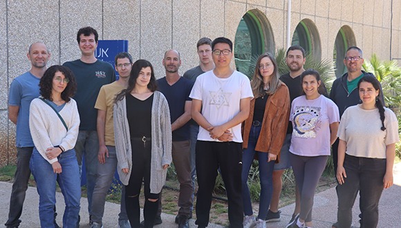
The research team
The team addressed this challenge by using CRISPR and designing sgRNA sequences that guide an enzyme called Cas9 found naturally in bacteria to cut specific genetic sequences associated with entire gene families. Prof. Mayrose explains that "this genetic editing method allows us to design different sgRNA sequences to allow Cas9 to cut almost any gene that we want to change. We wanted to apply this technique to improve the control of creating mutations in plants for the purposes of agricultural improvement, and specifically to overcome the common limitation posed by genetic redundancy."
In the first stage, a bioinformatics study was carried out on a computer, which, unlike most studies in the field, initially covered the entire genome. The researchers chose to focus on the Arabidopsis plant, which is used as a model in many studies and has about 30,000 genes. First, they identified and isolated about 8,000 individual genes, which have no family members, and therefore no copies in the genome. The remaining 22,000 genes were divided into families, and for each family appropriate sgRNA sequences were computationally designed. Each sgRNA sequence was designed to guide the Cas9 cutting enzyme to a specific genetic sequence that characterizes the entire family, with the aim of creating mutations in all family members so that these genes can no longer overlap each other. In this way, a library was built that totaled approximately 59,000 sgRNA sequences, where each sgRNA by itself can simultaneously modify 2-10 genes at once from each gene family, thereby effectively neutralizing the phenomenon of genetic redundancy.
In addition, the sgRNA sequences were divided into ten sub libraries of approximately 6,000 sgRNA sequences each, according to the presumed role of the genes – such as coding for enzymes, receptors, transcription factors, etc. According to the researchers, establishing the libraries allowed them to focus and optimize the search for genes responsible for desired traits, a search that until now has been largely random.
"We believe that this is the future of agriculture: controlled and targeted crop improvement on a large scale. Today, we are applying the method we developed to rice and tomato plants with great success, and we intend to apply it to other crops as well." – Prof. Eilon Shani
In the next step, the researchers moved from the computer to the laboratory. Here they generated all 59,000 sgRNA sequences designed by the computational method and engineered them into new plasmid libraries (i.e., circular DNA segments) in combination with the cutting enzyme. The researchers then generated thousands of new plants containing the libraries - where each plant was implanted with a single sgRNA sequence directed against a specific gene family.
The researchers observed the traits that were manifested in the plants following the genome modifications, and when an interesting phenotype was observed in a particular plant, it was easy to know which genes were responsible for the change based on the sgRNA sequence that was inserted into it. Also, through DNA sequencing of the identified genes, it was possible to determine the nature of the mutation that caused the change and its contribution to the plant's new properties.
In this way, many new traits were mapped that until now were blocked due to genetic redundancy. Specifically, the researchers identified specific proteins that comprise a mechanism related to the transport of the hormone cytokinin, which is essential for optimal plant development.
Prof. Shani concludes: "The new method we developed is expected to be of great help to basic research in understanding processes in plants, but beyond that, it has enormous significance for agriculture: it makes it possible to efficiently and accurately reveal the pool of genes responsible for traits we seek to improve - such as resistance to drought, pests, and diseases, or increasing yields. We believe that this is the future of agriculture: controlled and targeted crop improvement on a large scale. Today we are applying the method we developed to rice and tomato plants with great success, and we intend to apply it to other crops as well."
Recognizing the transformative potential of this breakthrough, Tel Aviv University's technology commercialization company, Ramot, partnered with the AgChimedes group to establish DisTree, a company dedicated to applying this technology to different crops. This collaboration, along with financial investment and professional support, aims to revolutionize agricultural genetics and ensure nutritional security in the face of climate crises.

Research
Deadly epidemic killed all the black sea urchins in the Gulf of Eilat, placing coral reefs at risk
Recent, unsettling studies conducted by Tel Aviv University have unveiled a deadly epidemic responsible for the widespread decimation of black sea urchins in the Mediterranean Sea and the Gulf of Eilat. Over the span of just a few months, the entire population of black sea urchins in Eilat was eradicated. For instance, within a few weeks, thousands of sea urchins inhabiting a site near the northern shore of the Gulf of Eilat perished. The severity of the epidemic is such that only skeletal remains of black urchins now occupy the site. Disturbingly, similar occurrences have been observed at various other locations in the Gulf of Eilat, as well as in neighboring countries including Jordan, Egypt, Saudi Arabia, Greece, and Turkey.
"At first we thought it was some kind of pollution or poisoning, or a local chemical spill (…) but when we examined additional sites in Eilat, Jordan, and Sinai, we quickly realized that this was not a local incident. All findings pointed to a rapidly spreading epidemic." - Dr. Omri Bronstein.
The studies were led by Dr. Omri Bronstein and PhD students Rotem Zirler, Lisa-Maria Schmidt, Gal Eviatar, and Lachan Roth from the School of Zoology, at The George S. Wise Faculty of Life Sciences, and The Steinhardt Museum of Natural History at Tel Aviv University. The papers were published in Frontiers in Marine science and Royal Society Open Science.
The researchers underscore the vital importance of sea urchins, particularly the long-spined Diadema setosum, as keystone species essential for the thriving equilibrium of coral reefs. They express a pressing concern, stating, "It must be understood that the threat to coral reefs is already at an all-time peak, and now a previously unknown variable has been added. This situation is unprecedented in the documented history of the Gulf of Eilat."
According to the researchers' hypothesis, the cause of the deadly epidemic can be attributed to a pathogenic ciliate parasite that has spread from the Mediterranean to the Red Sea. In response to the gravity of the situation, an urgent report outlining the current state has been submitted to the Israel Nature and Parks Authority, instigating deliberation on emergency measures to safeguard Israel's coral reefs.
"Sea urchins in general, and Diadema setosum in particular, are considered key species essential for the healthy functioning of coral reefs. The sea urchins are the reef's 'gardeners' – they feed on the algae and prevent them from taking over and suffocating the corals that compete with them for sunlight." – Dr. Omri Bronstein
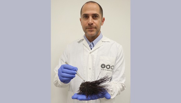
Dr. Omri Bronstein and a dying sea urchin
"At first we thought it was some kind of pollution or poisoning, or a local chemical spill, from the industry and hotels in the north of the Gulf of Eilat, but when we examined additional sites in Eilat, Jordan, and Sinai, we quickly realized that this was not a local incident," explains Dr. Bronstein. "All findings pointed to a rapidly spreading epidemic. Similar reports are coming in from colleagues in Saudi Arabia. Even sea urchins that we grow for research purposes in our aquariums at the Interuniversity Institute, and sea urchins at the Underwater Observatory Marine Park in Eilat, contracted the disease and died, probably because the pathogen got in through the pumping systems."
Dr. Bronstein describes it as a fast and violent death: "Within just two days a healthy sea urchin becomes a skeleton with massive tissue loss. While some corpses are washed ashore, most sea urchins are devoured while they are dying and unable to defend themselves, which could speed up contagion by the fish who prey on them."
In recent years, Dr. Bronstein's research group has dedicated their efforts to the investigation of marine invasions, with a specific focus on the long-spined Diadema setosum. "Until recently, the black sea urchins with long spines, familiar to many of us, was one of the dominant species in Eilat's coral reef," reflects Dr. Bronstein. "Sea urchins in general, and Diadema setosum in particular, are considered key species essential for the healthy functioning of coral reefs. The sea urchins are the reef's 'gardeners' – they feed on the algae and prevent them from taking over and suffocating the corals that compete with them for sunlight. Regrettably, these once-thriving sea urchins have vanished from the Gulf of Eilat and are quickly disappearing from constantly expanding parts of the Red Sea further to the south," shares Dr. Bronstein with a sense of lament.

A dying urchin in the Mediterranean Sea (photo: Dr. Omri Bronstein)
Several months ago, Dr. Bronstein was alerted to the initial reports of widespread mortality by colleagues in Greece and Turkey, where the sea urchins had invaded, likely via the Suez Canal. "In 2006, the first sighting of this species of sea urchin occurred in the southern regions of Turkey," Dr. Bronstein adds. This phenomenon, known as biological invasion, carries far-reaching ecological implications, pervasively affecting the eastern Mediterranean, particularly along Israel's coastline. "We have been monitoring the dynamics of this species' invasion in the Mediterranean since its first emergence," he shares.
In 2016, they discovered the first Diadema setosum sea urchin along Israel’s Mediterranean coastline – a lone urchin sighted at Gordon Beach in Tel Aviv. For over a decade since the first discovery in Turkey, the Mediterranean populations of these sea urchins remained small and usually hidden. However, since 2018 the sea urchin population in the Mediterranean has been growing exponentially, reaching a state of population explosion – with giant populations of thousands and even tens of thousands found in Greece and Turkey.
"The window of opportunity for preserving a thriving population of this species in Eilat has regrettably closed. To establish a safeguard population, we must act without delay, by preserving healthy individuals from the Israeli Mediterranean before the encroaching disease from the north reaches this region." Dr. Omri Bronstein
"However, during the course of our research, while scrutinizing the invasion of sea urchins in the Mediterranean, we began to receive reports on sudden extensive mortality," says Dr. Bronstein. "While the extinction of an invasive species is supposedly not a bad thing, we must be aware of two major risks: Firstly, we don't yet know how this mortality and its causes might impact local species in the Mediterranean. Secondly, and of far greater significance, the geographic proximity shared by the eastern Mediterranean and the Red Sea provides a potential conduit for the swift transmission of the pathogen into the Red Sea. As we feared and predicted, this is what appears to have happened."
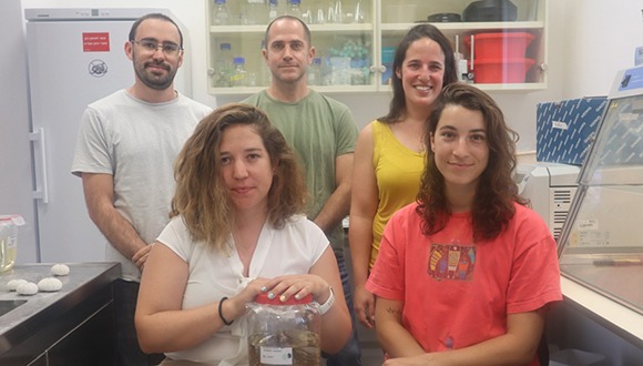
Dr. Bronstein and his research team (photo: courtesy of Dr. Omri Bronstein)
The massive loss of sea urchins reminded the TAU researchers of one of the most devastating events in marine ecology: the disappearance of the sea urchins in the Caribbean. Until 1983, the Caribbean coral reef thrived as a vibrant tropical ecosystem, much like the one in the Gulf of Eilat. But as the sea urchins vanished, the uncontrollable growth of algae took over, blocking sunlight from reaching the corals and forever altering the reef into a sea of algae.
Dr. Bronstein reveals, "Just last year, the Caribbean experienced another outbreak of the disease, resulting in the demise of the remaining urchin populations. However, unlike previous incidents, we now possess advanced scientific and technological resources to analyze the forensic evidence. Researchers from Cornell University successfully pinpointed the cause of mortality in the Caribbean: a pathogenic ciliate parasite. The identical pathology observed in the dying sea urchins of Greece, Turkey, and the Red Sea corroborates this finding."
Dr. Bronstein's pioneering research not only identified the unprecedented mass mortality of an invasive species in the Mediterranean but also shed light on the alarming decline of the widely prevalent sea urchin species, Diadema setosum. In a groundbreaking study, Dr. Bronstein issued a a warning that the epidemic plaguing the Mediterranean could extend its reach to the nearby Red Sea. Sadly, this cautionary prediction has become a disheartening reality.
"The gravity of the situation cannot be understated: the Red Sea is witnessing an alarming surge in mortality, surpassing the extent observed in the Mediterranean. Looming in the background is an ominous uncertainty: What is the exact cause of the sea urchin die-offs? Is it the same Caribbean pathogen or an entirely new and unfamiliar factor? Regardless, it is evident that this pathogen spreads through water, and we anticipate a rapid escalation of sickness and demise among the entire population of these sea urchins in both the Mediterranean and the Red Sea."
"In my view, it is imperative that we swiftly establish a safeguard population for these sea urchins, ensuring the potential for their reintroduction into the wild. Similar to the approach taken with COVID-19, the trajectory of this epidemic remains uncertain. Will it eventually subside on its own, or persist for years, radically transforming coral reefs? However, unlike the COVID-19 pandemic, there are no available vaccines or treatments for the afflicted sea urchins. Hence, our efforts must be steadfastly directed towards prevention. The window of opportunity for preserving a thriving population of this species in Eilat has regrettably closed. To establish a safeguard population, we must act without delay, by preserving healthy individuals from the Israeli Mediterranean before the encroaching disease from the north reaches this region. While this is a complex undertaking, it is imperative if we aspire to secure the future of this unique species, which plays a critical role in the destiny of coral reefs," concludes Dr. Bronstein.

