TAU researchers developed musical tests to detect mental deterioration in old age




Research
TAU researchers developed musical tests to detect mental deterioration in old age
Modern technology contributes to increased longevity and thus to the growth of the elderly population. It is therefore important to take steps to ensure their quality of life, including inventing tools for accessible and quick diagnosis of age-related conditions. While preventative tests are commonly accepted for a variety of physiological problems such as diabetes, high blood pressure or breast cancer, no method has yet been developed to enable routine, accessible monitoring of the brain for cognitive issues.
Researchers at Tel Aviv University have developed a method that employs musical tests and a portable instrument for measuring brain activity to detect cognitive decline in old age. The method entails measuring 15 minutes of electrical activity in the subject's brain while he or she performs simple musical tasks and can be easily implemented by any staff member in any clinic, without requiring special training. The researchers believe the method could pave the way towards early detection of cognitive decline when treatment and prevention of severe decline are possible, improving the quality of life of millions around the world
The study was led at Tel Aviv University by PhD student Neta Maimon from the School of Psychological Sciences and the Buchmann-Mehta School of Music, and Lior Molcho from Neurosteer Ltd, headed by Prof. Nathan Intrator from the Blavatnik School of Computer Science and the Sagol School of Neuroscience. Other participants included: Adi Sasson, Sarit Rabinowitz, and Noa Regev-Plotnick from the Dorot-Netanya Geriatric Medical Center. The article was published in the journal Frontiers in Aging Neuroscience.
As part of the study, the researchers developed a groundbreaking method combining a portable device for the measurement and innovative analysis of electroencephalography (EEG), developed by Neurosteer, and a short musical test of about 12-15 minutes, developed by Maimon.
"We have actually succeeded in illustrating that music is indeed an effective tool for measuring brain activity."
Maimon, who specializes in musical cognition, explains that music has great influence on different centers in the brain. On the one hand, music is known to be a quick mood stimulant, particularly of positive emotion. On the other hand, in different situations, music can be cognitively challenging, activating the frontal parts of the brain, especially if we try to concentrate on different aspects of the music, and at the same time perform a particular task. According to Maimon, if we combine these two capabilities, we can create cognitive tests that are quite complex, yet also pleasant and easy to perform.
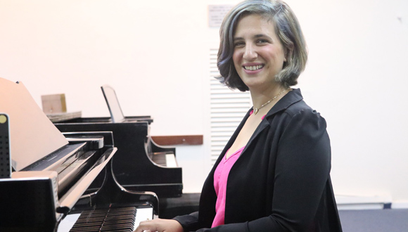
Neta Maimon specializes in musical cognition
Furthermore, music that is positive and reasonably rhythmic will enhance concentration and performance of the task. Thus, for example, the famous “Mozart effect,” whereby subjects perform better on intelligence tests after listening to Mozart’s music, has nothing to do with Mozart’s music, but rather the fact that music creates a positive mood and stimulates us to a state that is optimal for performing intelligence and creativity tests.
Accordingly, the researchers hypothesized that with musical tools, it would also be possible to challenge the subjects to an extent that would enable testing of the brain’s frontal activity as well as raising their spirits, thus enhancing their performance on the test while the overall experience is pleasant.
The study included an experiment testing 50 elderly people hospitalized at the Dorot-Netanya Geriatric Medical Center. “Anyone hospitalized at Dorot, or any other geriatric rehabilitation institution, undergoes a standard test called 'mini-mental,' designed to evaluate their cognitive condition as a routine part of the intake process," explains Maimon.
During the test, the subject is connected to the portable EEG device by means of an adhesive band with three electrodes attached to the forehead. The test includes a variety of tasks, including enumerating the days of the week or months of the year backwards. The subject performs a series of musical-cognitive tasks according to audible instructions given automatically through earphones. Short melodies are played by different instruments, and the subjects are instructed to perform various tasks on them at varying levels of difficulty. For example, pressing a button each time any melody is played or pressing it only when the violin plays. In addition, the test includes several minutes of musically guided meditation designed to bring the brain to a resting state, as this state is known to indicate cerebral functioning in various situations. Up to 30 points can be accrued, a high score indicating normal cognition.
"Our method enables the monitoring of cognitive capability and detection of cognitive decline already in the early stages, all by simple and accessible means."
"The participants scored 18-30 on the mini-mental test, indicating various levels of cognitive functioning," explains Maimon. "The EEG device registered the electrical activity in the brain during the activity, and the results were analyzed using machine learning technology. This allowed mathematical indices to be identified that were precisely correlated with the mini-mental test scores; in other words, we obtained new neuro-markers [brain markers] that may stand alone as indices of the subject’s cognitive status."
Maimon adds: “We have actually succeeded in illustrating that music is indeed an effective tool for measuring brain activity. The brain activity and response times to tasks correlated to the subjects’ cerebral conditions (correlating to the mini-mental score assigned to them). More importantly, all those who underwent the experiment reported that, on the one hand, it challenged the brain, but on the other it was very pleasant to perform”.
The researchers conclude: “Our method enables the monitoring of cognitive capability and detection of cognitive decline already in the early stages, all by simple and accessible means, with a quick and easy test that can be conducted in any clinic. This method is of special importance today due to the increase in longevity and accelerated population growth, particularly among the elderly. Today, millions of people around the world already suffer or are liable to suffer soon from cognitive decline and its dire consequences, and their number will only increase in the coming decades. Our method could pave the way towards efficient cognitive monitoring of the general population, and thus detect cognitive decline in its early stages, when treatment and prevention of severe decline are possible. It is therefore expected to improve the quality of life of millions around the world.”

Research
Scientific discovery may facilitate speedy, objective, and accurate diagnosis of the condition using saliva
A scientific breakthrough from the Tel Aviv and Haifa Universities may facilitate speedy, objective, and accurate diagnosis of people suffering from posttraumatic stress disorder, PTSD, using saliva samples, as well as developing microbiotic related medications (associated with the body's microbial ecology).
The study was a joint effort by eminent scholars from various fields. It was led by Professor Illana Gozes and included Professor Noam Shomron, Dr. Shlomo Sragovich and Ph.D. student Guy Shapira, (all from TAU’s Sackler Faculty of Medicine and Sagol School of Neuroscience) as well as Prof. Zahava Solomon from TAU’s Gershon H. Gordon Faculty of Social Sciences, and Prof. Abraham Sagi-Schwartz and PhD student Ella Levert-Levitt from the Center for the Study of Child Development and the School of Psychological Sciences at Haifa University. The study was published in NATURE's prestigious MOLECULAR PSYCHIATRY magazine.
The researchers tested a unique group of about 200 Israeli veteran soldiers (they all came from a larger cohort of subjects from a comprehensive four-decade-long study of veterans by Prof. Solomon) who had fought in the first Lebanon War in 1982. The test covered various psychological aspects, including sleep, appetite disorders, guilt, suicidal thoughts, social and spousal support, hostility, satisfaction with life, as well as issues of demographics, psychopathology, welfare, health, and education.
"We were surprised to discover that about a third of the PTSD subjects had never been diagnosed with post-trauma, so they never received any recognition from the Ministry of Defense and the official authorities."
The researcher also collected saliva samples from them and comparing the results of the subjects' microbial distribution to the psychological results and their responses to the welfare questionnaires, the researchers from the universities of Tel Aviv and Haifa found that people with PTSD and high psychopathological indications exhibit the same picture of bacteria in the saliva (a unique oral microbiotic signature).
According to the researchers, this study is significant in that for the first time, we might be able to diagnose post-trauma by objective criteria and not just behavioral ones.
It is interesting to note here that the saliva bacteria of those exposed to air pollution showed a correlation to the picture with PTSD, while the number of years of education showed a protective influence and a reverse picture of the microbial ecology in the saliva.
"To the best of our knowledge, this is the first depiction of a microbial signature in the saliva among veteran soldiers with PTSD," says Prof. Illana Gozes. "We were surprised to discover that about a third of the PTSD subjects had never been diagnosed with post-trauma, so they never received any recognition from the Ministry of Defense and the official authorities."
"To the best of our knowledge, this is the first depiction of a microbial signature in the saliva among veteran soldiers with PTSD,"
"It must be stressed that until now, post-trauma diagnosis has been based solely on psychological and psychiatric measures. Thanks to this study, it may be possible, in the future, to use objective molecular and biological characteristics to distinguish PSTD sufferers, taking into account environmental influences. We hope that this new discovery and the microbial signatures described in this study might promote easier diagnosis of post-traumatic veteran soldiers so they can receive appropriate treatment."
The study was also supported by IDF's Medical Corps Department of Health and Well-Being and Dr. Ariel Ben Yehuda, former chief of the above Department and currently, a Department Manager in the Mental Health Medical Center in Shalvata, Clallit Health Services. The study also involved collaboration with the Charité University Medicine in Berlin and its microbiology experts Dr. Markus M. Heimesaat and Professor Stefan Bereswill, as well as with the University of Hong Kong, which is studying the effects of air pollution, Professors Victor Li and Jacqueline Lam.

Research
Newly discovered molecule may allow for more accessible and effective cancer immunotherapies
Researchers at Tel Aviv University and the University of Lisbon have discovered a potentially new immunotherapy, which may lead to more affordable and effective treatments. Immunotherapy activates the patient’s immune system to fight cancer cells.
In this significant step in the fight against cancer, the researchers used computational and bioinformatic systems to discover a tiny molecule which can enter a solid tumor. Thanks to its low molecular weight, the molecule outperforms existing antibodies that are used as the key component in many cancer immunotherapies today. The molecule is also small enough that it may one day be administered in a pill form, saving stressful and time-consuming trips to the hospital.
Behind the groundbreaking development is an international team of researchers led by 2020 Kadar Family Award recipient Prof. Ronit Sachi-Fainaro, Director of the Center for Cancer Biology Research and Head of the Laboratory for Cancer Research and Nanomedicine at the Sackler Faculty of Medicine at Tel Aviv University, along with Prof. Helena Florindo and Prof. Rita Guedes from the Research Institute for Medicines at the Faculty of Pharmacy, University of Lisbon. The results of the study were published in the Journal for ImmunoTherapy of Cancer.
Immunotherapies can significantly improve patient recovery rates, without the severe side effects that accompany treatments such as chemotherapy. Immunotherapies often make use of antibodies, which are similar to proteins produced by the immune system to destroy infection-causing organisms. However, while lab-grown antibodies created to fight cancer have demonstrated some success, they are costly and not always effective.
“I believe that in the future, the small molecule will be commercially available and will make immunotherapy affordable for cancer patients."
Considering these challenges, TAU and University of Lisbon researchers used computational, bioinformatics and data analysis tools to evaluate thousands of molecular structures. They discovered a list of potential candidates and used the best structure they found to synthesize the new, small molecule which has successfully activated immune cells against cancer cells in lab models, including patient-derived ones.
The creation of this small molecule builds on the research of Nobel Prize winners James Allison and Tasuku Honjo, who originally developed the CTLA-4 and PD-1 antibodies, respectively, which are used in today's cancer immunotherapies. The two discovered that immune cells are essentially disabled by particular proteins found in cancer and immune cells. The protein called PD-L1 is found in cancer cells, and paralyzes immune cells by binding to a protein on these cells called PD-1. Honjo's antibodies neutralize the PD-1/PD-L1 protein bond, allowing the immune system to attack the cancer.
“Patients will probably be able to take it at home, orally, without the need for IV administration in the hospital."
Prof. Satchi-Fainaro, head of the TAU research team and a 2020 Kadar Family Award winner, explains that whereas lab-grown antibodies have complex structures and are expensive to produce, the new molecule was synthesized with simple equipment at a low cost. “I believe that in the future, the small molecule will be commercially available and will make immunotherapy affordable for cancer patients."
The small molecule is also better equipped to penetrate a solid tumor than previous treatments. The antibodies used for current treatments enter a tumor via its blood vessels. "If there are fewer blood vessels in a particular area of the tumor, the antibody will not be able to get inside. The small molecule, on the other hand, diffuses, and is therefore not entirely dependent on the tumor's blood vessels or on its hyper-permeability,” says Prof. Satchi-Fainaro. “Another advantage of the small molecule is that it may be available in a format that patients will probably be able to take at home, orally, without the need for intravenous injections in the hospital."
This work was supported by Fundação para a Ciência e a Tecnologia, Ministério da Ciência, Tecnologia e Ensino Superior (FCT-MCTES) and by The Israeli Ministry of Health under the frame of EuroNanoMed-II, “La Caixa” Foundation, Liga Portuguesa Contra o Cancro, the European Research Council (ERC), The Israel Science Foundation, The Melanoma Research Alliance (MRA), the Israel Cancer Research Fund (ICRF) Professorship award and the Morris Kahn Foundation.

Research
Could Accelerated Learning Process and Improve Visual Perception Capabilities?
A new study from Tel Aviv University proposes a new learning method for people with autism that may accelerate the learning process and significantly improve capabilities in terms of visual perception. According to the researchers, improving the perceptual capacity of people with autism is often a challenge, and usually requires long and tedious training alongside additional learning challenges that characterizes autism, such as the ability to generalize learning to new situations.
The study was conducted by doctoral student Shira Klorfeld-Auslender and Prof. Nitzan Censor from the School of Psychological Sciences and the Sagol School of Neuroscience at Tel Aviv University, in collaboration with Prof. Ilan Dinstein and his team from Ben-Gurion University. The study was published in the journal Current Biology.
"A large part of learning does not happen in formal training settings but afterwards, through processes of assimilation and reinforcement of memory that occur in an 'offline' state; for example, when our brain is asleep."
The new method proposed by the researchers is based on utilizing "memory flashes," by exposing a person for just a few seconds to a task that has already been learned. While standard teaching practice reinforce length and repetition of new skills, the new method improved both visual perception capabilities and the generalization of learning through helping the subjects excel in the same tasks, under different conditions.
"In my laboratory, we focus on the study of learning in humans, and we know that a large part of learning does not happen in formal training settings but afterwards, through processes of assimilation and reinforcement of memory that occur in an 'offline' state; for example, when our brain is asleep," explains Prof. Censor.
"However, standard teaching methods still advocate an approach where longer practice equals better learning: if you want to play the piano, you should practice playing the piano for many hours every day until the playing becomes second nature to you. We have identified an alternative learning mechanism that uses 'memory flashes' – a brief exposure to a task that has already been learned –to assimilate and generalize skill developed."
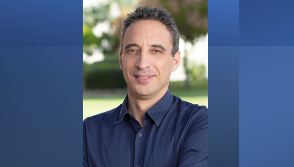
Prof. Nitzan Censor
In the study, 30 high-functioning adults with autism were asked to learn a visual task (for example, identifying the direction of lines that appear for a few milliseconds on the screen). However, instead of repeating the task for a long time each day, the examinees in the main experimental group learned the task in depth on the first day, and in the following days they were exposed to the visual stimulus for only a few seconds. At the end of the process, although the study participants studied the task for a minimal amount of time, their performance improved significantly, by about 20–25%, which was a similar result to those subjected to multiple-repetition learning and to the achievements of subjects without autism.
"We have shown that it does not take prolonged practice time to assimilate the task – it is enough to flash it for a few seconds to stimulate the relevant brain network, and the brain will then assimilate the material on its own."
Moreover, even when presented with the task under new conditions (for example, when the stimulus was learned in a new location), the examinees who learned with the memory flash method performed better than those in the control group - they knew how to generalize the skills learned in the first task. The participants' success in generalizing the learning to other situations is considered significant, as these are skills that people with autism tend to struggle with.
"We have already proven in previous studies that processes of learning assimilation can be improved through flashes of memory," says Prof. Censor. "We have shown that it does not take prolonged practice time to assimilate the task – it is enough to flash it for a few seconds to stimulate the relevant brain network, and the brain will then assimilate the material on its own."
"In this case, we tested people with autism. People with autism often have difficulty learning and generalizing repetitive learning, that is, using tools that have also been learned when executing new tasks. Through short flashes of visual stimulus of a task learned, we were able to produce learning that is identical to repetitive learning in terms of its effectiveness; meaning, we significantly shortened the learning time. The added value is the ability to generalize: the examinees performed a task under new conditions, as if they had fully learned it. "
According to Prof. Censor, the new method may have significant potential implications in a wide range of areas. The new study could pave the way for more meaningful approaches to learning for people with autism, and in a wide variety of tasks. Moreover, the method may contribute to shorten rehabilitation after neurological injuries.

Research
TAU researchers solve centuries old scientific mystery
For centuries, nature lovers and scientists have been fascinated by the fact that creatures in the sea are able to glow. The phenomenon is very common in reef-building corals, but its biological role has been the subject of constant debate. Numerous hypotheses have been tested over the years. Some suggested that this phenomenon protect against radiation. Or perhaps it contributed to the optimization of the photosynthesis? Maybe the glowing property helped protect the coral against herbivores or to attract symbiotic algae to the corals?
A new Tel Aviv University study, in collaboration with the Steinhardt Museum of Natural History, and the Interuniversity Institute for Marine Sciences in Eilat, has proven for the first time that the magical phenomenon – whereby corals in deep reefs display glowing colors (fluorescence) - is intended to serve as a mechanism for attracting prey.
The study was led by Dr. Or Ben-Zvi, in collaboration with Yoav Lindemann and Dr. Gal Eyal, under the supervision of Prof. Yossi Loya from the School of Zoology and the Steinhardt Museum of Natural History at Tel Aviv University.
The researchers first sought to determine whether plankton (small organisms that drift in the sea along with the current) are attracted to fluorescence, both in the laboratory and at sea. Then, in the lab, the researchers quantified the predatory capabilities of mesophotic corals (corals that live between the shallow coral reef area and the deep, completely dark zone of the ocean), which exhibit different fluorescent appearances.
To test the planktons’ potential attraction to fluorescence, the researchers used, among other things, the crustacean Artemia salina, which is used in many experiments as well as for food for corals. The researchers noted that when the crustaceans were given a choice between a green or orange, fluorescent target versus a clear 'control' target, they showed a significant preference for the fluorescent target.
Moreover, when the crustaceans were given a choice between two clear targets, its choices were observed to be randomly distributed in the experimental setup. In all of the laboratory experiments, the crustaceans vastly exhibited a preferred attraction toward a fluorescent signal. Similar results were presented when using a native crustacean from the Red Sea. However, unlike the crustaceans, fish that are not considered coral prey did not exhibit these trends, and rather avoided the fluorescent targets.

The second phase of the study was carried out about 40 meters deep in the sea, where the fluorescent traps (both green and orange) attracted twice as many plankton as the clear trap.
“We conducted an experiment in the depths of the sea to examine the possible attraction of diverse and natural collections of plankton to fluorescence, under the natural currents and light conditions that exist in deep water," says Dr. Or Ben-Zvi. "Since fluorescence is ‘activated’ principally by blue light (the light of the depths of the sea), at these depths the fluorescence is naturally illuminated, and the data that emerged from the experiment were unequivocal, similar to the laboratory experiment.”
"This phenomenon may play a greater role in marine ecosystems than previously thought."
In the last part of the study, the researchers examined the predation rates of mesophotic corals that were collected at 45 m depth in the Gulf of Eilat. They found that corals that displayed green fluorescence enjoyed predation rates that were 25 percent higher than corals exhibiting yellow fluorescence.
Prof. Loya: “Many corals display a fluorescent color pattern that highlights their mouths or tentacle tips, a fact that supports the idea that fluorescence, like bioluminescence (the production of light by a chemical reaction), acts as a mechanism to attract prey. The study proves that the glowing and colorful appearance of corals can act as a lure to attract swimming plankton to ground-dwelling predators, such as corals, and especially in habitats where corals require other energy sources in addition or as a substitute for photosynthesis (sugar production by symbiotic algae inside the coral tissue using light energy).”
Dr. Ben-Zvi concludes: “Despite the gaps in the existing knowledge regarding the visual perception of fluorescence signals by plankton, the current study presents experimental evidence for the prey-luring role of fluorescence in corals. We suggest that this hypothesis, which we term the ‘light trap hypothesis’, may also apply to other fluorescent organisms in the sea, and that this phenomenon may play a greater role in marine ecosystems than previously thought.”

Research
TAU Researchers caution that while the genome editing method is very effective, it is not always safe and can promote cancer
A new study from TAU identifies risks in the use of CRISPR therapeutics – an innovative, Nobel-prize-winning method that involves cleaving and editing DNA, already employed for the treatment of conditions like cancer, liver and intestinal diseases, and genetic syndromes.
Investigating the impact of this technology on T-cells (white blood cells of the immune system), the researchers detected a loss of genetic material in a significant percentage – up to 10% of the treated cells. They explain that such loss can lead to destabilization of the genome, which might cause cancer.
The study was led by Dr. Adi Barzel from the School of Neurobiology, Biochemistry and Biophysics at TAU's George S. Wise Faculty of Life Sciences and Dotan Center for Advanced Therapies, a collaboration between the Tel Aviv Sourasky Medical Center (Ichilov) and Tel Aviv University, and by Dr. Asaf Madi and Dr. Uri Ben-David from TAU's Sackler Faculty of Medicine and Edmond J. Safra Center for Bioinformatics. The findings were published in the leading scientific journal Nature Biotechnology.
The researchers explain that CRISPR is a groundbreaking technology for editing DNA – cleaving DNA sequences at certain locations to delete unwanted segments, or alternately repair or insert beneficial segments. Developed about a decade ago, the technology has already proved impressively effective in treating a range of diseases – cancer, liver diseases, genetic syndromes, and more.
The first approved clinical trial ever to use CRISPR, was conducted in 2020 at the University of Pennsylvania, when researchers applied the method to T-cells – white blood cells of the immune system. Taking T-cells from a donor, they expressed an engineered receptor targeting cancer cells, while using CRISPR to destroy genes coding for the original receptor – which otherwise might have caused the T-cells to attack cells in the recipient's body.
"CRISPR therapeutics, in which DNA is cleaved intentionally as a means for treating cancer, might, in extreme scenarios, actually promote malignancies."
In the present study, the researchers sought to examine whether the potential benefits of CRISPR therapeutics might be offset by risks resulting from the cleavage itself, assuming that broken DNA is not always able to recover.
Dr. Ben-David and his research associate Eli Reuveni explain, "The genome in our cells often breaks due to natural causes, but usually it is able to repair itself, with no harm done. Still, sometimes a certain chromosome is unable to bounce back, and large sections, or even the entire chromosome, are lost. Such chromosomal disruptions can destabilize the genome, and we often see this in cancer cells. Thus, CRISPR therapeutics, in which DNA is cleaved intentionally as a means for treating cancer, might, in extreme scenarios, actually promote malignancies."
To examine the extent of potential damage, the researchers repeated mentioned 2020 Pennsylvania experiment, cleaving the T-cells' genome in the same locations – chromosomes 2, 7, and 14 (of the human genome's 23 pairs of chromosomes). Using a state-of-the-art technology called 'single-cell RNA sequencing' they analyzed each cell separately and measured the expression levels of each chromosome in every cell.
A significant loss of genetic material was detected in some of the cells. For example, when Chromosome 14 had been cleaved, about 5% of the cells showed little or no expression of this chromosome. When all chromosomes were cleaved simultaneously, the damage increased, with 9%, 10%, and 3% of the cells unable to repair the break in chromosomes 14, 7, and 2 respectively. The three chromosomes did differ, however, in the extent of the damage they sustained.
"Single-cell RNA sequencing and computational analyses enabled us to obtain very precise results," explain Dr. Madi and his student Ella Goldschmidt, adding: "We found that the cause for the difference in damage was the exact place of the cleaving on each of the three chromosomes. Altogether, our findings indicate that over 9% of the T-cells genetically edited with the CRISPR technique had lost a significant amount of genetic material. Such loss can lead to destabilization of the genome, which might promote cancer."
"We advance this highly effective technology, while at the same time cautioning against its potential dangers. This may seem like a contradiction, but as scientists we are quite proud of our approach, because we believe that this is the very essence of science: we don't 'choose sides.'"
Based on their findings, the researchers caution that extra care should be taken when using CRISPR therapeutics. They also propose alternative, less risky, methods, for specific medical procedures, and recommend further research into two kinds of potential solutions: reducing the production of damaged cells or identifying damaged cells and removing them before the material is administered to the patient.
Dr. Barzel and his PhD student Alessio Nahmad conclude: "Our intention in this study was to shed light on potential risks in the use of CRISPR therapeutics. We did this even though we are aware of the technology's substantial advantages. In fact, in other studies we have developed CRISPR-based treatments, including a promising therapy for AIDS. We have even established two companies – one using CRISPR and the other deliberately avoiding this technology. In other words, we advance this highly effective technology, while at the same time cautioning against its potential dangers. This may seem like a contradiction, but as scientists we are quite proud of our approach, because we believe that this is the very essence of science: we don't 'choose sides.' We examine all aspects of an issue, both positive and negative, and look for answers."