


A new study from Tel Aviv University’s Moshe Dayan Center finds major shifts in political attitudes among Arab citizens of Israel, including record-high support for an Arab party joining the next governing coalition.


Research
Joint study by Tel Aviv University, the IDF Medical Corps, and the U.S. Department of Defense confirms and strengthens earlier findings
A joint study by Tel Aviv University, the IDF Medical Corps, and the U.S. Department of Defense has found that a series of specialized computer-based training exercises can significantly reduce the risk of post-traumatic stress disorder (PTSD) among IDF combat soldiers. The new study confirmed and extends similar results obtained in a comparable trial conducted in 2012. Unfortunately, the program that was created as a result of this earlier study and implemented by the IDF in 2018 was shut down shortly before the outbreak of the Iron Swords War following budget cuts and restructuring in the army’s Mental Health Department.
The new study, involving 500+ soldiers from one of the IDF’s regular infantry brigades, was conducted in 2022–2023 (before the outbreak of the war) under the leadership of Prof. Yair Bar-Haim, Director of the National Center for Traumatic Stress and Resilience and member of the School of Psychological Sciences at Tel Aviv University, together with doctoral student Chelsea Gober Dykan. The results now appear in the American Journal of Psychiatry, one of the world’s leading journals in the field.
Prof. Bar-Haim explains that the computer-based training involves a simple task in which soldiers are shown both neutral and threatening images or words that are replaced by target shapes that appear on the screen near those stimuli. The soldiers are asked to identify the targets, a process that gradually trains them to direct more attention toward potential threats in their environment. Each session lasts about ten minutes and is completed individually, four times over separate days.
The earlier study, conducted in 2012, tested the effectiveness of the training among roughly 800 soldiers in one of the IDF’s infantry brigades. The soldiers completed the computerized training during basic training. In the summer of 2014, during Israel’s Operation Protective Edge, most of those soldiers saw combat. Four months after the fighting ended, Prof. Bar-Haim and his team found that 7.8% of the soldiers who had not received the training were diagnosed with PTSD, compared with just 2.6% among those who completed the training program.
Prof. Bar-Haim, Chelsea Gober Dykan, and their research team recently set out to replicate the earlier findings and reevaluate the training’s effectiveness, this time using slightly modified computer-based protocols. This study was conducted in 2022–2023 (before the outbreak of the Iron Swords War) at the brigade training base during advanced training. One-third of the soldiers received the original attentional training protocol, another third underwent a revised attentional training protocol, and the remaining third received placebo training. After completing their training, the soldiers were deployed on their first operational rotation in Judea and Samaria. Upon their return, the researchers assessed the impact of the different training protocols on the soldiers’ risk for developing PTSD.
The results were once again clear-cut. Among soldiers in the control (placebo) group, 5.3% reported clinically significant post-traumatic symptoms, compared to 2.7% in the group that received the revised attention-training protocol, and less than one percent (0.9%) among those who completed the original attention training version. The results show that the original protocol remained the most effective in reducing the risk of PTSD, while the revised method demonstrated lower efficacy.
“Replication of findings is a critical component of clinical science and provides confidence in the validity of results,” explains Prof. Bar-Haim. “We once again found that the attentional training we developed is effective in reducing the risk of PTSD among soldiers deployed on operational settings, which further strengthens our confidence in its impact - that’s the good news. However, we also saw that the additional method we tested, based on eye-tracking technology, proved to be less effective. That’s how it is in science: our hypotheses don’t always hold up under rigorous testing, and we therefore must draw conclusions accordingly and refine our tools through further research.”
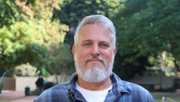
Prof. Yair Bar-Haim, Head of the National Center for Traumatic Stress and Resilience
Prof. Bar-Haim notes that the less encouraging news is that the program was discontinued several months before the outbreak of the war, and thus was not available in its most potent and tested form to soldiers heading into the Gaza and Lebanon campaigns. To still provide some form of support ahead of the ground maneuvers in Gaza and Lebanon, the researchers, together with the IDF, developed a mobile application that allows soldiers to complete the training on their personal phones rather than at designated computer stations. The app, called “Combat Attention” (Keshev Kravi), was distributed to both regular forces and reservists prior to the start of the ground operation.
According to Prof. Bar-Haim, the new findings not only reinforce confidence in attentional training as an effective tool for building resilience and preventing PTSD among combat soldiers, but also can serve as a wake-up call to military decision-makers to reinstate the program at brigade training bases and expand its use across combat units.
Prof. Bar-Haim concludes: “The study was conducted before the war when soldiers’ operational activities where typical to the low-intensity combat experiences at that time. The results demonstrated significant differences in PTSD risk between soldiers who underwent the computer-based training and those who did not, rendering the program valuable during routine deployments. It is likely that in wartime, these gaps become even more pronounced, making attentional training even more desirable. During wartime, armies typically reach their peak operational capabilities, and the same often holds true for mental health care and prevention efforts. Now, as the war subsides, the greatest challenge for the IDF is to preserve and sustain these hard-won capabilities, ensuring they are not lost in quieter times when their importance may seem less urgent. Decision-makers are urged to act now to allocate the necessary budgets and design long-term evidence-based solutions for PTSD prevention and mitigation among deploying troops.

Research
TAU meta-analysis finds pathogens, storms, and extreme temperatures are the leading causes of sea urchin mass mortality events.
Two pioneering studies by researchers from the School of Zoology and the Steinhardt Museum of Natural History at Tel Aviv University, led by Dr. Omri Bronstein, have identified the primary drivers of sea urchin mass mortality events over recent decades: pathogens, storms, and extreme temperatures. In addition, Dr. Bronstein and his team have developed an innovative method for genetic sampling in marine environments - using a swab similar to a COVID-19 test — to enable rapid and non-invasive monitoring of marine animals and underwater disease outbreaks.
The first study, published in the journal Biological Reviews, presents a meta-analysis of all 110 scientifically documented mass mortality events (MMEs) among sea urchins recorded between 1888 and 2024. Dr. Bronstein and PhD student Lisa Schmidt conducted a comprehensive review of the history of these events, showing that most reported MMEs originate in the Northern Hemisphere — particularly in the United States, Western Europe, and Japan — where the majority of research and funding are concentrated. The Tel Aviv University researchers classified five main causes of these events and found that 33% were caused by pathogens, 25% by catastrophic events such as storms and oxygen depletion, 24% by extreme temperatures, 11% by algal blooms, and 7% by human activity, such as pollution and habitat destruction.
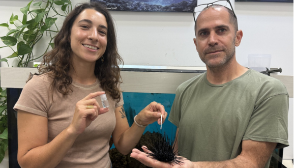
Left to right: Mai Bonomo & Dr. Omri Bronstein holding sea urchin and sample tube
“This is a meta-analysis of all scientific literature on the subject,” says Dr. Bronstein. “For each mass mortality event, we mapped where and when it occurred, which species were affected, and most importantly — what the causes were. After filtering out hundreds of publications who lacked sufficient credible data to be included in our analyses, ee found that pathogens are the leading cause of mass mortalities among sea urchins. This finding aligns closely with what we are seeing today in the modern wave of die-offs — from the Caribbean to the Red Sea and the Indian Ocean. There is a tendency to attribute everything to global warming, but that is not always accurate. In many cases, mortality is not directly related to heat, as some affected sea urchin species naturally live in even warmer environments. These temperatures may not be optimal, but they are not lethal for these species. The problem is that warming influences many other environmental factors, which can combine into a deadly mix. For example, warmer waters tend to have lower dissolved oxygen and higher pathogen activity.”
In 2023, Dr. Bronstein identified a mass mortality event of long-spined sea urchins (Diadema setosum) along the Red Sea coast. He subsequently found that the same pathogen — a ciliate parasite — responsible for wiping out a related Caribbean species was also to blame. Since that discovery, the outbreak has spread to the Indian Ocean, reappeared in the Caribbean, and is now considered a global pandemic threatening sea urchin populations worldwide.
“Sea urchins are vital to coral reef health,” explains Dr. Bronstein. “They are the ‘gardeners’ of the reef: they feed on algae and prevent it from overgrowing and suffocating the corals competing for sunlight. In 1983, the most dominant Caribbean sea urchin species, Diadema antillarum, died in vast numbers from an unknown reason at the time; algae proliferated uncontrollably, shaded the corals, and the entire ecosystem shifted from coral reefs to algal fields. Even 40 years later, the sea urchin population — and the reefs — have not recovered. We fear that the same process may now occur in other parts of the world where mass die-offs are happening, mainly among the long-spined sea urchin, a relative of the Caribbean species — the black urchin with long spines familiar to everyone. Until recently, it was one of the most common reef urchins in Eilat; today it has almost disappeared from large parts of the Red Sea. This is a very violent event: within less than 48 hours, a healthy population turns into disintegrating skeletons. In some sites in Eilat and Sinai, mortality reached 100%. Later, mass deaths were recorded on Réunion Island in the Indian Ocean, and we are now investigating three additional mass mortality events in the Atlantic and Indian Ocean, and even the Mediterranean Seas. What began as a local mortality event has become regional and then global, posing a threat to coral reefs everywhere.”
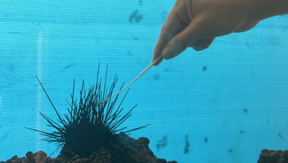
Close-up of hand swabbing sea urchin underwater tank
To address one of the major challenges in marine genetic sampling, graduate student Mai Bonomo and Dr. Bronstein published a separate study in Molecular Ecology Resources, developing a new, inexpensive, and non-invasive method for collecting underwater genetic samples at scale.
“The main tools used today to identify both animals and pathogens are genetic,” says Dr. Bronstein. “But molecular ecology faces a fundamental problem: there’s no simple way to sample DNA from live marine animals underwater. As a result, many studies rely on invasive methods that harm the animal or even require sacrificing it completely to bring it into the lab. Therefore, research in this field is heavily regulated, weighing each case’s scientific value against environmental ethics. For example, sampling is prohibited in marine nature reserves, there are restrictions and bans on shipping samples abroad — including corals — and every scientific publication must present the official permits for each sample it reports. Our need to overcome this bottleneck arose from the sea urchin pandemic. Today, there are only two ways to detect diseased urchins: visually — which is too late, as the animals are already dying — or through genetic tools that can detect disease before symptoms appear. But if detecting disease requires removing the animal from the sea, it makes no difference whether it’s sick or not — we end up sacrificing it.”
To overcome this challenge, Tel Aviv University researchers developed a specialized underwater genetic sampling kit that is durable, reliable, inexpensive, and easy to use — and it is already being adopted by research groups worldwide, especially in remote or sensitive areas.
“We developed a new tool for underwater DNA sampling that resembles a COVID-19 test,” explains Dr. Bronstein. “At the end of a special tube filled with a preservation liquid is a membrane preventing water penetration, sealed with a clip-cap — much like some toothpaste tubes. Just like a COVID test, the researcher gently swabs the surface of the marine animal, without harming or moving it. There’s no need to collect mucus as in humans — just a light swipe is enough. The swab is then inserted into the tube, piercing the membrane that protects the preservation liquid inside, and the cap is locked to secure the sample. That’s it. A single researcher can collect dozens of samples in one dive, under almost any environmental or depth conditions.
The kit has already been tested in challenging environments, including field expeditions to Djibouti and Réunion Island, and the results are very promising: samples remained exceptionally well-preserved for months without refrigeration before arriving at our lab, and still allowed for sensitive genetic analyses. In a large-scale trial we conducted in the Gulf of Eilat, we collected genetic material from hundreds of echinoderms — the group that includes sea urchins and starfish — within just a few months, and performed the most extensive genetic analysis ever conducted on these species in the region. This led to the discovery of several new species and the reclassification of others previously unknown to science. This is a simple and elegant solution to one of the most persistent technical challenges in marine molecular ecology.”

Research
TAU researchers identify a key molecular mechanism behind ALS and succeed in stopping, and even reversing, nerve degeneration.
A new international study led by Tel Aviv University researchers may pave the way for an effective treatment for amyotrophic lateral sclerosis (ALS), a fatal and currently incurable neurodegenerative disease.
The team uncovered a previously unknown molecular mechanism that drives the progression of ALS and succeeded in neutralizing it using RNA-based gene therapy. “When we added a specific RNA molecule to human cells and animal models for ALS, the nerve cells stopped degenerating and even regenerated,” the researchers said. The breakthrough findings may offer new hope to millions of patients worldwide.
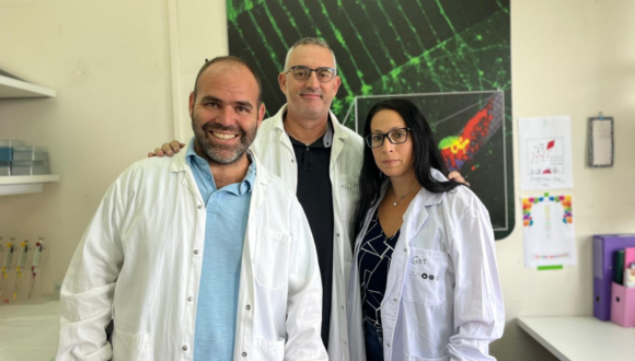
The research team (Left to right): Dr. Ariel Ionescu, Prof. Eran Perlson & Tal Pery Gradus.
The study was conducted in the laboratory of Prof. Eran Perlson from the Gray Faculty of Medical & Health Sciences and the Sagol School of Neuroscience at Tel Aviv University. It was led by Dr. Ariel Ionescu and Dr. Lior Ankol, in collaboration with Dr. Amir Dori, Senior Neurologist and Head of the Neuromuscular Disease Unit at Sheba Medical Center. Additional participants included researchers from the Weizmann Institute of Science, Ben-Gurion University of the Negev, and research institutions in France, Turkey, and Italy. The paper was published in the leading neuroscience journal Nature Neuroscience.
Prof. Perlson explains: “Our lab studies ALS - a fatal, incurable neurodegenerative disease. ALS affects motor neurons and causes gradual paralysis of all muscles in the body. Most patients die within 3–5 years of diagnosis, due to paralysis of the diaphragm muscles and respiratory failure. We know that in ALS, the neuromuscular junctions - where nerve fibers (axons) meet muscle cells and transmit electrical signals from the brain to the muscles — are disrupted. However, the molecular mechanisms causing this damage remained unknown until now, and consequently no effective treatment has been developed. In this study, we wanted to get to the root of the matter and generate new knowledge that would enable the development of effective drugs for ALS.”

Illustration showing the gene therapy’s protective effect on motor neurons, preventing the “fire-like” degeneration characteristic of ALS
The current study was based on a feature of ALS discovered previously in Prof. Perlson’s lab: toxic clusters (aggregates) of a protein called TDP-43 (usually responsible for regulating protein production at the site) form at the tip of the nerve, where it meets the muscle. To discover how these TDP-43 aggregates are formed, the researchers used a mouse model for ALS, tissues from ALS patients, and cultures of human stem cells.
The study found that muscle cells produce small RNA molecules called microRNA-126 and send them in vesicles, through the synapsis, to the tip of the nerve cell. The role of these molecules is to prevent the expression of the TDP-43 protein at the neuromuscular junction when it is not needed. Dr. Ionescu explains: “We discovered that in ALS, the muscle produces a smaller amount of microRNA-126, which leads to an excess of TDP-43. The excess protein forms toxic aggregates that attack molecules essential for functioning of the mitochondria — the nerve cell’s powerhouse. Damage to the mitochondria causes an energy deficit, gradually destroying motor neurons and leaving patients’ muscles paralyzed.”
The study further showed that when the amount of microRNA-126 is reduced, a process similar to ALS occurs, and the neurons are destroyed. Conversely, increasing the level of microRNA-126 in tissues taken from ALS patients and in ALS model mice led to a decrease in the levels of TDP-43, and the neurons stopped degenerating and even regenerated. The researchers concluded that adding microRNA-126 rescues neurons damaged by ALS, prevents degeneration of the neuromuscular junction, and could serve as a basis for developing effective drugs for this currently incurable disease.
Prof. Perlson concludes: “In this study, we identified for the first time a critical molecular mechanism of ALS in its early stages: a reduction in the amount of microRNA-126 transferred from muscle to nerve, resulting in the formation of toxic aggregates of the TDP-43 protein that kill neurons. Our findings may serve as a basis for developing an effective gene therapy focused on adding microRNA-126, which could bring hope to millions of patients and their families around the world.”

Research
A new TAU study reveals how the insurance sector, one of the world’s largest financial forces, can take a leading role in the global response to climate change.
As global warming intensifies and extreme weather events become more frequent, insurance systems worldwide are under pressure. In the United States, for instance, rising flood and hurricane damages have driven major reforms in the federal flood insurance program (NFIP), reducing public subsidies and raising costs for homeowners.
Published in Humanities and Social Sciences Communications (Nature Portfolio), the study examines how climate change–driven hurricanes could impact profitability in the U.S. homeowners’ insurance market — and proposes a new approach: transforming anticipated financial losses into climate-mitigation investments.
The research was conducted by a joint team from Tel Aviv University, Max Stern Yezreel Valley College, and the University of Haifa, including PhD student Moran Nabriski and Prof. Colin Price from TAU’s Porter School of the Environment and Earth Sciences, and Dr. Ruslana Palatnik from the University of Haifa.
Insurance is a major economic force with a dual role; on the one hand it is a risk manager, and on the other a large institutional investor with long-duration capital. Given its systemic weight – and because insurance is fundamentally a pooling mechanism that links economic sectors – the study calls for the industry to be a proactive partner in addressing climate change. It should not only react to extreme events but also reduce risk at its source (akin to building-safety standards that prevent fire losses).
Insurance plays a dual role in the global economy: it manages risk and serves as a powerful institutional investor. Given its influence and financial reach, the researchers argue that the industry should act not only as a responder to natural disasters, but as a proactive force in reducing climate risks, much like building codes prevent fire losses before they occur.
By combining a market-equilibrium model with climate-driven hurricane damage projections, the study shows that insurers’ profitability could decline by 11%–100% across modeled scenarios, leading to higher premiums and reduced coverage. Redirecting that expected loss into emissions-reduction initiatives, the researchers note, could generate climate benefits that far exceed the industry’s direct economic share.
“Insurance is commonly viewed as a tool for transferring risk over time and across geographies, yet natural disasters occur in the same places at the same time,” said lead author Moran Nabriski. “As natural disasters intensify, the insurance industry should represent the economy not only as a responder to a changing climate, but also as a leader in confronting it. Because insurance connects all sectors of the economy, it can leverage that position into a coordinated effort with a meaningful impact on climate risk.”
The study provides a quantitative framework for assessing future risks and demonstrates how insurers’ long-term capital can become a powerful engine for financing global climate solutions.


 teams at the signing of the cleanroom manufacturing agreement.")