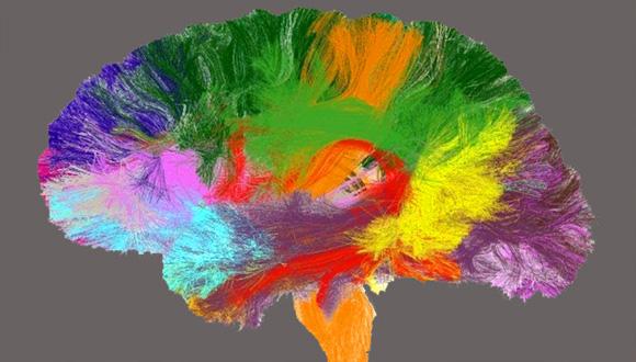
TAU-led project to create brain ‘atlas’ hailed a success
New map of the brain will help improve the accuracy and efficiency of research and treatment for neurological diseases
Researchers from Tel Aviv University recently led a joint project – called CONNECT – in cooperation with researchers from all over Europe. CONNECT was comprised of twelve groups of researchers and attempted to create the first comprehensive "atlas" of the microstructures in the white matter of the human brain. The project relied on MRI technology, and received 2.5 million euros in funding from the European Union, under the category of 'technologies of the future'. The study's participants were located in leading research centers in Israel, England, Germany, France, Denmark, Switzerland and Italy. On Friday, October 19th, after three years of collaborative research, they met in Paris to announce the successful completion of the project.
The human brain in 3D
"Today medical researchers use an old atlas of the brain, based on the mind of one particular man who donated his body to science," says Prof. Yaniv Assaf of the Department of Neurobiology and Sagol School of Neuroscience at Tel Aviv University. Prof. Assaf, who also heads TAU's Alfredo Federico Strauss Center for Computational Neuro-Imaging, was the initiator of the international project and served as its coordinator. "The new atlas, on the other hand, is based MRI samples from many subjects, and is even presented in 3D. Because the methodology employed in this project, as well as its scope, the new atlas is far more detailed and accurate. It lets us examine the brain in a way that, prior to this study, was only possible with a microscope."
The first major contribution of the new atlas is its mapping of white matter – which is composed of nerve fibers that transmit information – in the brains of living humans rather than the subjects of autopsies. This breakthrough was made possible with the use of MRI technology, which allows scientists to map the brain using non-invasive methods. The second major contribution is the fact that the atlas is based on the scans of 120 healthy subjects ages 25-35, instead of data from just one person. This provides a broad and accurate picture of the normal structure of the human brain.
The new atlas presents microstructures in the brain in an organized, coded way that makes the data accessible even to professionals who don't specialize in brain research, such as doctors and researchers from other fields. Images represent various parameters, collected and measured using MRI, such as fiber thickness and density in different areas of the brain.
From gray matter to white matter
In addition to its medical contribution, the project is expected to significantly advance the study of white matter in the brain, its activities and functions. "Until now, neuroscientists have focused mainly on so-called 'gray matter,' says Prof. Assaf. "They did not investigate the 'white matter,' which is about half the volume of the brain, mainly because they lacked effective research tools. The MRI method we developed will allow researchers to observe, for the first time, what goes on in living brain tissue. It will introduce a whole new world of possibility."
Prof. Assaf and his colleagues are also planning to use technology to examine the dynamics and evolution over time of microstructures in white matter. For example, they'll be looking for the "fingerprints" left behind by cognitive processes, such as learning a new piece of information. Another goal of their research is developing a better understanding of the changes various diseases, such as Alzheimer's or schizophrenia, bring about in the brain, to help develop better diagnostic methods.


