Visualizing Brainpower
In an ambitious, international research initiative spearheaded by TAU's Prof. Yaniv Assaf (pictured below), Head of the Strauss Center for Computational Neuroimaging, scientists used MRI technology to scan the brains of 120 healthy people to create the first complete “atlas” of white matter. This spongy, whitish nerve tissue makes up about 50 percent of the brain’s volume and is responsible for transmitting information between neurons. The European Union-funded project, known as CONNECT, is providing insight into the microstructure of the human brain.

"The roadmap we developed will enable researchers to compare healthy brains to sick ones," says Prof. Assaf, a member of the George. S. Wise Faculty of Life Sciences and Sagol School of Neuroscience. "This could eventually assist in early detection and treatment of neurological and psychiatric diseases.” While in the past, scientists mapped the brain by analyzing the organs of cadavers under a microscope, Assaf’s team used noninvasive MRI techniques – developed at the Strauss Center – to create 3D images of the brains of living Israelis and Europeans for the CONNECT project.
Pioneering neuroimaging
Since the advent of neuroimaging in Israel in the mid-1990s, Tel Aviv University has been at the forefront of scientific advancement in this interdisciplinary field.For example, using unique MRI tracking methods at the Strauss Center's animal model facility, Prof. Yoram Cohen of the Ramond and Beverly Sackler School of Chemistry transplanted stem cells gleaned from bone marrow into the brain. Initial results indicate that the stem cells can identify unhealthy or damaged tissues, migrate to them and potentially repair or halt cell degeneration.
In a separate study using the animal MRI, Prof. Ina Weiner of the School of Psychological Sciences found that if you give schizophrenia patients drug treatments in early adolescence, you can prevent the devastating effects of the disorder. The animal MRI scanner has been the basis for more than 30 papers in major scientific journals, as well as the research of over a dozen graduate students.
Next year, the Center will become equipped with a state-of-the-art human MRI. This new facility is expected to contribute to important developments in our understanding of human behavior and neurological diseases. About 20 teams from 5 faculties will use the powerful MRI to explore what they were unable to see before: images of activity in various parts of the human brain.
Zooming in on a single neuron
PhD candidate Daniel Barazany (pictured below), an avid photographer and tech-lover, is developing sophisticated neuroimaging tools at the Strauss Center. He is searching for the “holy grail” of neuroscience: what the brain looks like and how it works on the level of a single nerve cell. While a conventional MRI scanner can capture a general image of the brain, it is not able to zoom in on the brain’s smallest units. As a result, present understanding of the basic building blocks of the brain is incomplete.
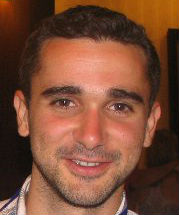
In collaboration with Prof. Assaf, Daniel uses MRI tools to image, in detail, microscopic layers of nerve cells. “If we could study the brain’s microstructure up close," he explains, "we could potentially pinpoint the precise moment abnormalities occur. With this knowledge, we could develop drugs that target specific neurons and treat devastating neurodegenerative diseases such as ALS and MS.”
A recipient of a Colton Scholarship, Daniel is currently involved in five different MRI experiments at the Strauss Center. He collaborates with Prof. Galit Yovel at the School of Psychological Sciences to study the areas of the brain responsible for vision, and with Prof. Amir Amedi from Hebrew University to examine differences in the microstructure of blind peoples’ brains. He also collaborates and shares ideas with his wife Hilit Levy, another neuroscience doctoral student at the Department of Neurobiology.
The interdisciplinary nature of neuroscience is the main reason Daniel decided to get involved in the field: “When you study one scientific field, you get one opinion. Neuroscience combines physics, chemistry, biology, medicine, psychology, art, engineering and more. You are exposed to so many different schools of thought.” Daniel hopes to one day combine all of his interests and start a career in the neuroscience industry.

